Tuesday, 22 February 2011 17:05

Lung Function Examination

Rate this item
(3 votes)

Lung function may be measured in a number of ways. However, the aim of the measurements has to be clear before the examination, in order to interpret the results correctly. In this article we will discuss lung function examination with special regard to the occupational field. It is important to remember the limitations in different lung function measurements. Acute temporary lung function effects may not be discernible in case of exposure to fibrogenic dust like quartz and asbestos, but chronic effects on lung function after long-term (>20 years) exposure may be. This is due to the fact that chronic effects occur years after the dust is inhaled and deposited in the lungs. On the other hand, acute temporary effects of organic and inorganic dust, as well as mould, welding fumes and motor exhaust, are well suited to study. This is due to the fact that the irritative effect of these dusts will occur after a few hours of exposure. Acute or chronic lung function effects also may be discernible in cases of exposure to concentrations of irritating gases (nitrogen dioxide, aldehydes, acids and acid chlorides) in the vicinity of well documented exposure limit values, especially if the effect is potentiated by particulate air contamination.

Lung function measurements have to be safe for the examined subjects, and the lung function equipment has to be safe for the examiner. A summary of the specific requirements for different kinds of lung function equipment are available (e.g., Quanjer et al. 1993). Of course, the equipment must be calibrated according to independent standards. This may be difficult to achieve, especially when computerized equipment is being used. The result of the lung function test is dependent on both the subject and the examiner. To provide satisfactory results from the examination, technicians have to be well trained, and able to instruct the subject carefully and also encourage the subject to carry out the test properly. The examiner should also have knowledge about the airways and lungs in order to interpret the results from the recordings correctly.

It is recommended that the methods used have a fairly high reproducibility both between and within subjects. Reproducibility may be measured as the coefficient of variation, that is, the standard deviation multiplied by 100 divided by the mean value. Values below 10% in repeated measurements on the same subject are deemed acceptable.

In order to determine if the measured values are pathological or not, they must be compared with prediction equations. Usually the prediction equations for spirometric variables are based on age and height, stratified for sex. Men have on the average higher lung function values than women, of the same age and height. Lung function decreases with age and increases with height. A tall subject will therefore have higher lung volume than a short subject of the same age. The outcome from prediction equations may differ considerably between different reference populations. The variation in age and height in the reference population will also influence the predicted values. This means, for example, that a prediction equation must not be used if age and/or height for the examined subject are outside the ranges for the population that is the basis for the prediction equation.

Smoking will also diminish lung function, and the effect may be potentiated in subjects who are occupationally exposed to irritating agents. Lung function used to be considered as not being pathological if the obtained values are within 80% of the predicted value, derived from a prediction equation.

Measurements

Lung function measurements are carried out to judge the condition of the lungs. Measurements may either concern single or multiple measured lung volumes, or the dynamic properties in the airways and lungs. The latter is usually determined through effort-dependent manoeuvres. The conditions in the lungs may also be examined with regard to their physiological function, that is, diffusion capacity, airway resistance and compliance (see below).

Measurements concerning ventilatory capacity are obtained by spirometry. The breathing manoeuvre is usually performed as a maximal inspiration followed by a maximal expiration, vital capacity (VC, measured in litres). At least three technically satisfactory recordings (i.e., full inspiration and expiration effort and no observed leaks) should be done, and the highest value reported. The volume may be directly measured by a water-sealed or a low-resistive bell, or indirectly measured by pneumotachography (i.e., integration of a flow signal over time). It is important here to note that all measured lung volumes should be expressed in BTPS, that is, body temperature and ambient pressure saturated with water vapour.

Forced expired vital capacity (FVC, in litres) is defined as a VC measurement performed with a maximally forced expiratory effort. Due to the simplicity of the test and the relatively inexpensive equipment, the forced expirogram has become a useful test in the monitoring of lung function. However, this has resulted in many poor recordings, of which the practical value is debatable. In order to carry out satisfactory recordings, the updated guideline for the collection and use of the forced expirogram, published by the American Thoracic Society in 1987, may be useful.

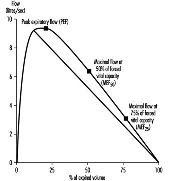
Instantaneous flows may be measured on flow-volume or flow-time curves, while time average flows or times are derived from the spirogram. Associated variables which can be calculated from the forced expirogram are forced expired volume in one second (FEV1, in litres per second), in percentage of FVC (FEV1%), peak flow (PEF, l/s), maximal flows at 50% and 75% of forced vital capacity (MEF50 and MEF25, respectively). An illustration of the derivation of FEV1 from the forced expirogram is outlined in figure 1. In healthy subjects, maximal flow rates at large lung volumes (i.e., at the beginning of expiration) reflect mainly the flow characteristics of the large airways while those at small lung volumes (i.e., the end of expiration) are usually held to reflect the characteristics of the small airways, figure 2. In the latter the flow is laminar, while in the large airways it may be turbulent.

Figure 1. Forced expiratory spirogram showing the derivation of FEV1 and FVC according to the extrapolation principle.

RES030F1


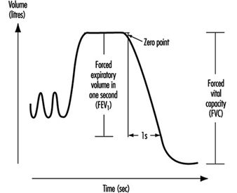
Figure 2.  Flow-volume curve showing the derivation of peak expiratory flow (PEF), maximal flows at 50% and 75% of forced vital capacity (and , respectively).

RES030F2

PEF may also be measured by a small portable device such as the one developed by Wright in 1959. An advantage with this equipment is that the subject may carry out serial measurements—for example, at the workplace. To get useful recordings, however, it is necessary to instruct the subjects well. Moreover, one should keep in mind that measurements of PEF with, for example, a Wright meter and those measured by conventional spirometry should not be compared due to the different blow techniques.

The spirometric variables VC, FVC and FEV1 show a reasonable variation between individuals where age, height and sex usually explain 60 to 70% of the variation. Restrictive lung function disorders will result in lower values for VC, FVC and FEV1. Measurements of flows during expiration show a great individual variation, since the measured flows are both effort and time dependent. This means, for example, that a subject will have extremely high flow in case of diminished lung volume. On the other hand, the flow may be extremely low in case of very high lung volume. However, the flow is usually decreased in case of a chronic obstructive disease (e.g., asthma, chronic bronchitis).

Figure 3.  A principal outline of the equipment for determination of total lung capacity (TLC) according to the helium dilution technique.

RES030F3

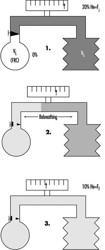
The proportion of residual volume (RV), that is, the volume of air which still is in the lungs after a maximal expiration, can be determined by gas dilution or by body plethysmography. The gas dilution technique requires less complicated equipment and is therefore more convenient to use in studies carried out at the workplace. In figure 3, the principle for the gas dilution technique has been outlined. The technique is based on dilution of an indicator gas in a rebreathing circuit. The indicator gas must be sparingly soluble in biological tissues so that it is not taken up by the tissues and blood in the lung. Hydrogen was initially used, but because of its ability to form explosive mixtures with air it was replaced by helium, which is easily detected by means of the thermal conductivity principle.

The subject and the apparatus form a closed system, and the initial concentration of the gas is thus reduced when it is diluted into the gas volume in the lungs. After equilibration, the concentration of indicator gas is the same in the lungs as in the apparatus, and functional residual capacity (FRC) can be calculated by means of a simple dilution equation. The volume of the spirometer (including the addition of the gas mixture into the spirometer) is denoted by VS, VL is the volume of the lung, Fi is the initial gas concentration and Ff is the final concentration.

FRC = VL = [(VS · Fi) / Ff] – VS


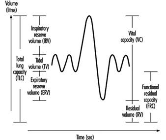
Two to three VC manoeuvres are carried out to provide a reliable base for the calculation of TLC (in litres). The subdivisions of the different lung volumes are outlined in figure 4.


Figure 4. Spirogram labelled to show the subdivisions of the total capacity.

RES030F4

Due to change in the elastic properties of the airways, RV and FRC increase with age. In chronic obstructive diseases, increased values of RV and FRC are usually observed, while VC is decreased. However, in subjects with badly ventilated lung areas—for example, subjects with emphysema—the gas dilution technique may underestimate RV, FRC and also TLC. This is due to the fact that the indicator gas will not communicate with closed-off airways, and therefore the decrease in the indicator gas concentration will give erroneously small values.


Figure 5. A principal outline of the recording of airway closure and the slope of the alveolar plateau (%).

RES030F5

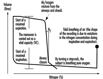
Measures of airway closure and gas distribution in the lungs can be obtained in one and the same manoeuvre by the single breath wash-out technique, figure 5. The equipment consists of a spirometer connected to a bag-in-box system and a recorder for continuous measurements of nitrogen concentration. The manoeuvre is carried out by means of a maximal inspiration of pure oxygen from the bag. In the beginning of the expiration, the nitrogen concentration increases as a result of emptying the subject’s deadspace, containing pure oxygen. The expiration continues with the air from the airways and alveoli. Finally, air from the alveoli, containing 20 to 40% nitrogen, is expired. When the expiration from the basal parts of the lungs increases, the nitrogen concentration will rise abruptly in case of airway closure in dependent lung regions, figure 5. This volume above RV, at which airways close during an expiration, is usually expressed as closing volume (CV) in percentage of VC (CV%). Distribution of the inspired air in the lungs is expressed as the slope of the alveolar plateau (%N2 or phase III, %N2/l). It is obtained by taking the difference in nitrogen concentration between the point when 30% of the air is exhaled and the point for airway closure, and dividing this by the corresponding volume.

Ageing as well as chronic obstructive disorders will result in increased values for both CV% and phase III. However, not even healthy subjects have a uniform gas distribution in the lungs, resulting in slightly elevated values for phase III, that is, 1 to 2% N2/l. The variables CV% and phase III are considered to reflect the conditions in the peripheral small airways with an internal diameter about 2 mm. Normally, the peripheral airways contribute to a small part (10 to 20%) of the total airway resistance. Quite extensive changes which are not detectable by conventional lung function tests like dynamic spirometry, may occur, for example, as a result of an exposure to irritating substances in the air in the peripheral airways. This suggests that airway obstruction begins in the small airways. Results from studies also have shown alterations in CV% and phase III before any changes from the dynamic and static spirometry have occurred. These early changes may go into remission when exposure to hazardous agents has ceased.

The transfer factor of the lung (mmol/min; kPa) is an expression of the diffusion capacity of oxygen transport into the pulmonary capillaries. The transfer factor can be determined using single or multiple breath techniques; the single breath technique is considered to be most suitable in studies at the workplace. Carbon monoxide (CO) is used since the back pressure of CO is very low in the peripheral blood, in contrast to that of oxygen. The uptake of CO is assumed to follow an exponential model, and this assumption can be used to determine the transfer factor for the lung.

Determination of TLCO (transfer factor measured with CO) is carried out by means of a breathing manoeuvre including a maximal expiration, followed by a maximal inspiration of a gas mixture containing carbon monoxide, helium, oxygen and nitrogen. After a breath-holding period, a maximal exhalation is done, reflecting the content in the alveolar air, Figure 10. Helium is used for the determination of the alveolar volume (VA). Assuming that the dilution of CO is the same as for helium, the initial concentration of CO, before the diffusion has started, can be calculated. TLCO is calculated according to the equation outlined below, where k depends on the dimensions of the component terms, t is the effective time for breath-holding and log is base 10 logarithm. Inspired volume is denoted Vi and the fractions F of CO and helium are denoted by i and a for inspired and alveolar, respectively.

TLCO = k Vi (Fa,He/Fi,He) log (Fi,CO Fa,He/Fa,CO Fi,He) (t)-1


Figure 6. A principal outline of the recording of transfer factor

RES030F6

The size of TLCO will depend on a variety of conditions—for example, the amount of available haemoglobin, the volume of ventilated alveoli and perfused lung capillaries and their relation to each other. Values for TLCO decrease with age and increase with physical activity and increased lung volumes. Decreased TLCO will be found in both restrictive and obstructive lung disorders.

Compliance (l/kPa) is a function, inter alia, of the elastic property of the lungs. The lungs have an intrinsic tendency to collaborate—that is, to collapse. The power to keep the lungs stretched will depend on the elastic lung tissue, the surface tension in the alveoli, and the bronchial musculature. On the other hand, the chest wall tends to expand at lung volumes 1 to 2 litres above the FRC level. At higher lung volumes, power has to be applied to further expand the chest wall. At the FRC level, the corresponding tendency in the lungs is balanced by the tendency to expand. The FRC level is therefore denoted by the resting level of the lung.

The compliance of the lung is defined as the change in volume divided by the change in transpulmonary pressure, that is, the difference between the pressures in the mouth (atmospheric) and in the lung, as the result of a breathing manoeuvre. Measurements of the pressure in the lung are not easily carried out and are therefore replaced by measurements of the pressure in the oesophagus. The pressure in the oesophagus is almost the same as the pressure in the lung, and it is measured with a thin polyethylene catheter with a balloon covering the distal 10 cm. During inspiratory and expiratory manoeuvres, the changes in volume and pressure are recorded by means of a spirometer and pressure transducer, respectively. When the measurements are performed during tidal breathing, dynamic compliance can be measured. Static compliance is obtained when a slow VC manoeuvre is carried out. In the latter case, the measurements are carried out in a body plethysmograph, and the expiration is intermittently interrupted by means of a shutter. However, measurements of compliance are cumbersome to perform when examining exposure effects on lung function at the worksite, and this technique is considered to be more appropriate in the laboratory.

A decreased compliance (increased elasticity) is observed in fibrosis. To cause a change in volume, large changes in pressure are required. On the other hand, a high compliance is observed, for example, in emphysema as the result of loss of elastic tissue and therefore also elasticity in the lung.

The resistance in the airways essentially depends on the radius and length of the airways but also on air viscosity. The airway resistance (RL in (kPa/l) /s), can be determined by use of a spirometer, pressure transducer and a pneumotachograph (to measure the flow). The measurements may also be carried out using a body plethysmograph to record the changes in flow and pressure during panting manoeuvres. By administration of a drug intended to cause broncho-constriction, sensitive subjects, as a result of their hyperreactive airways, may be identified. Subjects with asthma usually have increased values for RL.

Acute and Chronic Effects of Occupational Exposure on Pulmonary Function

Lung function measurement may be used to disclose an occupational exposure effect on the lungs. Pre-employment examination of lung function should not be used to exclude job-seeking subjects. This is because the lung function of healthy subjects varies within wide limits and it is difficult to draw a borderline below which it can safely be stated that the lung is pathological. Another reason is that the work environment should be good enough to allow even subjects with slight lung function impairment to work safely.

Chronic effects on the lungs in occupationally exposed subjects may be detected in a number of ways. The techniques are designed to determine historical effects, however, and are less suitable to serve as guidelines to prevent lung function impairment. A common study design is to compare the actual values in exposed subjects with the lung function values obtained in a reference population without occupational exposure. The reference subjects may be recruited from the same (or nearby) workplaces or from the same city.

Multivariate analysis has been used in some studies to assess differences between exposed subjects and matched unexposed referents. Lung function values in exposed subjects may also be standardized by means of a reference equation based on lung function values in the unexposed subjects.

Another approach is to study the difference between the lung function values in exposed and unexposed workers after adjustment for age and height with the use of external reference values, calculated by means of a prediction equation based on healthy subjects. The reference population may also be matched to the exposed subjects according to ethnic group, sex, age, height and smoking habits in order to further control for those influencing factors.

The problem is, however, to decide if a decrease is large enough to be classified as pathological, when external reference values are being used. Although the instruments in the studies have to be portable and simple, attention must be paid both to the sensitivity of the chosen method for detecting small anomalies in airways and lungs and the possibility of combining different methods. There are indications that subjects with respiratory symptoms, such as exertion dyspnoea, are at a higher risk of having an accelerated decline in lung function. This means that the presence of respiratory symptoms is important and so should not be neglected.

The subject may also be followed-up by spirometry, for example, once a year, for a number of years, in order to give a warning against the development of illness. There are limitations, however, since this will be very time-consuming and the lung function may have deteriorated permanently when the decrease can be observed. This approach therefore must not be an excuse for delay in carrying out measures in order to decrease harmful concentrations of air pollutants.

Finally, chronic effects on lung function may also be studied by examining the individual changes in lung function in exposed and unexposed subjects over a number of years. One advantage of the longitudinal study design is that the intersubject variability is eliminated; however, the design is considered to be time-consuming and expensive.

Susceptible subjects may also be identified by comparing their lung function with and without exposure during working shifts. In order to minimize possible effects of diurnal variations, lung function is measured at the same time of day on one unexposed and one exposed occasion. The unexposed condition can be obtained, for example, by occasionally moving the worker to an uncontaminated area or by use of a suitable respirator during a whole shift, or in some cases by performing lung function measurements in the afternoon of a worker’s day off.

One special concern is that repeated, temporary effects can result in chronic effects. An acute temporary lung function decrease may not only be a biological exposure indicator but also a predictor of a chronic lung function decrement. Exposure to air pollutants may result in discernible acute effects on lung function, although the mean values of the measured air pollutants are below the hygienic limit values. The question thus arises, whether these effects really are harmful in the long run. This question is hard to answer directly, especially since the air pollution in workplaces often has a complex composition and the exposure cannot be described in terms of mean concentrations of single compounds. The effect of an occupational exposure is also partly due to the sensitivity of the individual. This means that some subjects will react sooner or to a larger extent than others. The underlying pathophysiological ground for an acute, temporary decrease in lung function is not fully understood. The adverse reaction upon exposure to an irritating air contaminant is, however, an objective measurement, in contrast to subjective experiences like symptoms of different origin.

The advantage of detecting early changes in airways and lungs caused by hazardous air pollutants is obvious—the prevailing exposure may be reduced in order to prevent more severe illnesses. Therefore, an important aim in this respect is to use the measurements of acute temporary effects on lung function as a sensitive early warning system that can be used when studying groups of healthy working people.

Monitoring of Irritants

Irritation is one of the most frequent criteria for setting exposure limit values. It is, however, not certain that compliance with an exposure limit based on irritation will protect against irritation. It should be considered that an exposure limit for an air contaminant usually contains at least two parts—a time-weighted average limit (TWAL) and a short-term exposure limit (STEL), or at least rules for exceeding the time-weighted average limit, “excursion limits”. In the case of highly irritating substances, such as sulphur dioxide, acrolein and phosgene, it is important to limit the concentration even during very short periods, and it has therefore been common practice to fix occupational exposure limit values in the form of ceiling limits, with a sampling period that is kept as short as the measuring facilities will allow.

Time-weighted average limit values for an eight-hour day combined with rules for excursion above these values are given for most of the substances in the American Conference of Governmental Industrial Hygienists (ACGIH) threshold limit value (TLV) list. The TLV list of 1993-94 contains the following statement concerning excursion limits for exceeding limit values:

“For the vast majority of substances with a TLV-TWA, there is not enough toxicological data available to warrant a STEL = short-term exposure limit). Nevertheless, excursions above the TLV-TWA should be controlled even where the eight-hour TWA is within recommended limits.”

Exposure measurements of known air contaminants and comparison with well documented exposure limit values should be carried out on a routine basis. There are, however, many situations when the determination of compliance with exposure limit values is not enough. This is the case in the following circumstances (inter alia):

  1. when the limit value is too high to safeguard against irritation
  2. when the irritant is unknown
  3. when the irritant is a complex mixture and there is no suitable indicator known.


As advocated above, the measurement of acute, temporary effects on lung function can be used in these cases as a warning against over-exposure to irritants.

In cases (2) and (3), acute, temporary effects on lung function may be applicable also in testing the efficiency of control measures to decrease exposure to air contamination or in scientific investigations, for example, in attributing biological effects to components of air contaminants. A number of examples follow in which acute, temporary lung function effects have been successfully employed in occupational health investigations.

Studies of Acute, Temporary Lung Function Effects

Work-related, temporary decrease of lung function over a work shift was recorded in cotton workers at the end of 1950. Later, several authors reported work-related, acute, temporary changes of lung function in hemp and textile workers, coal miners, workers exposed to toluene di-isocyanate, fire-fighters, rubber processing workers, moulders and coremakers, welders, ski waxers, workers exposed to organic dust and irritants in water-based paints.

However, there are also several examples where measurements before and after exposure, usually during a shift, have failed to demonstrate any acute effects, despite a high exposure. This is probably due to the effect of normal circadian variation, mainly in lung function variables depending on the size of airway calibre. Thus the temporary decrease in these variables must exceed the normal circadian variation to be recognized. The problem may be circumvented, however, by measuring lung function at the same time of the day at each study occasion. By using the exposed employee as his or her own control, the interindividual variation is further decreased. Welders were studied in this way, and although the mean difference between unexposed and exposed FVC values was less than 3% in 15 examined welders, this difference was significant at the 95% confidence level with a power of more than 99%.

The reversible transient effects on the lungs can be used as an exposure indicator of complicated irritating components. In the study cited above, particles in the work environment were crucial for the irritating effects on the airways and lungs. The particles were removed by a respirator consisting of a filter combined with a welding helmet. The results indicated that the effects on the lungs were caused by the particles in welding fumes, and that the use of a particulate respirator might prevent this effect.

Exposure to diesel exhaust also gives measurable irritative effects on the lungs, shown as an acute, temporary lung function decrease. Mechanical filters mounted on the exhaust pipes of trucks used in loading operations by stevedores relieved subjective disorders and reduced the acute, temporary lung function decrease observed when no filtration was done. The results thus indicate that the presence of particles in the work environment does play a role in the irritative effect on airways and lungs, and that it is possible to assess the effect by measurements of acute changes in lung function.

A multiplicity of exposures and a continually changing work environment may present difficulties in discerning the causal relationship of the different agents existing in a work environment. The exposure scenario in sawmills is an illuminating example. It is not possible (e.g., for economical reasons) to carry out exposure measurements of all possible agents (terpenes, dust, mould, bacteria, endotoxin, mycotoxins, etc.) in this work environment. A feasible method may be to follow the development of lung function longitudinally. In a study of sawmill workers in the wood-trimming department, lung function was examined before and after a working week, and no statistically significant decrease was found. However, a follow-up study carried out a few years later disclosed that those workers who actually had a numerical decrease in lung function during a working week also had an accelerated long-term decline in lung function. This may indicate that vulnerable subjects can be detected by measuring changes in lung function during a working week.


Back

Read 11521 times Last modified on Saturday, 23 July 2022 19:46

" DISCLAIMER: The ILO does not take responsibility for content presented on this web portal that is presented in any language other than English, which is the language used for the initial production and peer-review of original content. Certain statistics have not been updated since the production of the 4th edition of the Encyclopaedia (1998)."

Contents

Respiratory System References

Abramson, MJ, JH Wlodarczyk, NA Saunders, and MJ Hensley. 1989. Does aluminum smelting cause lung disease? Am Rev Respir Dis 139:1042-1057.

Abrons, HL, MR Peterson, WT Sanderson, AL Engelberg, and P Harber. 1988. Symptoms, ventilatory function, and environmental exposures in Portland cement workers. Brit J Ind Med 45:368-375.

Adamson, IYR, L Young, and DH Bowden. 1988. Relationship of alveolar epithelial injury and repair to the indication of pulmonary fibrosis. Am J Pathol 130(2):377-383.

Agius, R. 1992. Is silica carcinogenic? Occup Med 42: 50-52.

Alberts, WM and GA Do Pico. 1996. Reactive airways dysfunction syndrome (review). Chest 109:1618-1626.
Albrecht, WN and CJ Bryant. 1987. Polymer fume fever associated with smoking and use of a mold release spray containing polytetraflouroethylene. J Occup Med 29:817-819.

American Conference of Governmental Industrial Hygienists (ACGIH). 1993. 1993-1994 Threshold Limit Values and Biological Exposure Indices. Cincinnati, Ohio: ACGIH.

American Thoracic Society (ATS). 1987 Standards for the diagnosis and care of patients with chronic obstructive pulmonary disease (COPD) and asthma. Am Rev Respir Dis 136:225-244.

—.1995. Standardization of Spirometry: 1994 update. Amer J Resp Crit Care Med 152: 1107-1137.

Antman, K and J Aisner. 1987. Asbestos-Related Malignancy. Orlando: Grune & Stratton.

Antman, KH, FP Li, HI Pass, J Corson, and T Delaney. 1993. Benign and malignant mesothelioma. In Cancer: Principles and Practice of Oncology, edited by VTJ DeVita, S Hellman, and SA Rosenberg. Philadelphia: JB Lippincott.
Asbestos Institute. 1995. Documentation center: Montreal, Canada.

Attfield, MD and K Morring. 1992. An investigation into the relationship between coal workers’ pneumoconiosis and dust exposure in US coal miners. Am Ind Hyg Assoc J 53(8):486-492.

Attfield, MD. 1992. British data on coal miners’ pneumoconiosis and relevance to US conditions. Am J Public Health 82:978-983.

Attfield, MD and RB Althouse. 1992. Surveillance data on US coal miners’ pneumoconiosis, 1970 to 1986. Am J Public Health 82:971-977.

Axmacher, B, O Axelson, T Frödin, R Gotthard, J Hed, L Molin, H Noorlind Brage, and M Ström. 1991. Dust exposure in coeliac disease: A case-referent study. Brit J Ind Med 48:715-717.

Baquet, CR, JW Horm, T Gibbs, and P Greenwald. 1991. Socioeconomic factors and cancer incidence among blacks and whites. J Natl Cancer Inst 83: 551-557.

Beaumont, GP. 1991. Reduction in airborne silicon carbide whiskers by process improvements. Appl Occup Environ Hyg 6(7):598-603.

Becklake, MR. 1989. Occupational exposures: Evidence for a causal association with chronic obstructive pulmonary disease. Am Rev Respir Dis. 140: S85-S91.

—. 1991. The epidemiology of asbestosis. In Mineral Fibers and Health, edited by D Liddell and K Miller. Boca Raton: CRC Press.

—. 1992. Occupational exposure and chronic airways disease. Chap. 13 in Environmental and Occupational Medicine. Boston: Little, Brown & Co.

—. 1993. In Asthma in the workplace, edited by IL Bernstein, M Chan-Yeung, J-L Malo and D Bernstein. Marcel Dekker.

—. 1994. Pneumoconioses. Chap. 66 in A Textbook of Respiratory Medicine, edited by JF Murray and J Nadel. Philadelphia: WB Saunders.

Becklake, MR and B Case. 1994. Fibre burden and asbestos-related lung disease: Determinants of dose-response relationships. Am J Resp Critical Care Med 150:1488-1492.

Becklake, MR. et al. 1988. The relationships between acute and chronic airways responses to occupational exposures. In Current Pulmonology. Vol. 9, edited by DH Simmons. Chicago: Year Book Medical Publishers.

Bégin, R, A Cantin, and S Massé. 1989. Recent advances in the pathogenesis and clinical assessment of mineral dust pneumoconioses: Asbestosis, silicosis and coal pneumoconiosis. Eur Resp J 2:988-1001.

Bégin, R and P Sébastien. 1989. Alveolar dust clearance capacity as determinant of individual susceptibility to asbestosis: Experimental oservations. Ann Occup Hyg 33:279-282.

Bégin, R, A Cantin, Y Berthiaume, R Boileau, G Bisson, G Lamoureux, M Rola-Pleszczynski, G Drapeau, S Massé, M Boctor, J Breault, S Péloquin, and D Dalle. 1985. Clinical features to stage alveolitis in asbestos workers. Am J Ind Med 8:521-536.

Bégin, R, G Ostiguy, R Filion, and S Groleau. 1992. Recent advances in the early diagnosis of asbestosis. Sem Roentgenol 27(2):121-139.

Bégin, T, A Dufresne, A Cantin, S Massé, P Sébastien, and G Perrault. 1989. Carborundum pneumoconiosis. Chest 95(4):842-849.

Beijer L, M Carvalheiro, PG Holt, and R Rylander. 1990. Increased blood monocyte procoagulant activity in cotton mill workers. J. Clin Lab Immunol 33:125-127.

Beral, V, P Fraser, M Booth, and L Carpenter. 1987. Epidemiological studies of workers in the nuclear industry. In Radiation and Health: The Biological Effects of Low-Level Exposure to Ionizing Radiation, edited by R Russell Jones and R Southwood. Chichester: Wiley.

Bernstein, IL, M Chan-Yeung, J-L Malo, and D Bernstein. 1993. Asthma in the Workplace. Marcel Dekker.

Berrino F, M Sant, A Verdecchia, R Capocaccia, T Hakulinen, and J Esteve. 1995. Survival of Cancer Patients in Europe: The EUROCARE Study. IARC Scientific Publications, no 132. Lyon: IARC.

Berry, G, CB McKerrow, MKB Molyneux, CE Rossiter, and JBL Tombleson. 1973. A study of the acute and chronic changes in ventilatory capacity of workers in Lancashire Cotton Mills. Br J Ind Med 30:25-36.

Bignon J, (ed.) 1990. Health-related effects of phyllosilicates. NATO ASI series Berlin: Springer-Verlag.

Bignon, J, P Sébastien, and M Bientz. 1979. Review of some factors relevant to the assessment of exposure to asbestos dusts. In The use of Biological Specimens for the Assessment of Human Exposure to Environmental Pollutants, edited by A Berlin, AH Wolf, and Y Hasegawa. Dordrecht: Martinus Nijhoff for the Commission of the European Communities.

Bignon J, J Peto and R Saracci, (eds.) 1989. Non-occupational exposure to mineral fibres. IARC Scientific Publications, no 90. Lyon: IARC.

Bisson, G, G Lamoureux, and R Bégin. 1987. Quantitative gallium 67 lung scan to assess the inflammatory activity in the pneumoconioses. Sem Nuclear Med 17(1):72-80.

Blanc, PD and DA Schwartz. 1994. Acute pulmonary responses to toxic exposures. In Respiratory Medicine, edited by JF Murray and JA Nadel. Philadelphia: WB Saunders.

Blanc, P, H Wong, MS Bernstein, and HA Boushey. 1991. An experimental human model of a metal fume fever. Ann Intern Med 114:930-936.

Blanc, PD, HA Boushey, H Wong, SF Wintermeyer, and MS Bernstein. 1993. Cytokines in metal fume fever. Am Rev Respir Dis 147:134-138.

Blandford, TB, PJ Seamon, R Hughes, M Pattison, and MP Wilderspin. 1975. A case of polytetrafluoroethylene poisoning in cockatiels accompanied by polymer fume fever in the owner. Vet Rec 96:175-178.

Blount, BW. 1990. Two types of metal fume fever: mild vs. serious. Milit Med 155:372-377.

Boffetta, P, R Saracci, A Anderson, PA Bertazzi, Chang-Claude J, G Ferro, AC Fletcher, R Frentzel-Beyme, MJ Gardner, JH Olsen, L Simonato, L Teppo, P Westerholm, P Winter, and C Zocchetti. 1992. Lung cancer mortality among workers in the European production of man-made mineral fibers-a Poisson regression analysis. Scand J Work Environ Health 18:279-286.

Borm, PJA. 1994. Biological markers and occupational lung dsease: Mineral dust-induced respiratory disorders. Exp Lung Res 20:457-470.

Boucher, RC. 1981. Mechanisms of pollutant induced airways toxicity. Clin Chest Med 2:377-392.

Bouige, D. 1990. Dust exposure results in 359 asbestos-using factories from 26 countries. In Seventh International Pneumoconiosis Conference Aug 23-26, 1988. Proceedings Part II. Washington, DC: DHS (NIOSH).

Bouhuys A. 1976. Byssinosis: Scheduled asthma in the textile industry. Lung 154:3-16.

Bowden, DH, C Hedgecock, and IYR Adamson. 1989. Silica-induced pulmonary fibrosis involves the reaction of particles with interstitial rather than alveolar macrophages. J Pathol 158:73-80.

Brigham, KL and B Mayerick. 1986. Endotoxin and Lung injury. Am Rev Respir Dis 133:913-927.

Brody, AR. 1993. Asbestos-induced lung disease. Environ Health Persp 100:21-30.

Brody, AR, LH Hill, BJ Adkins, and RW O’Connor. 1981. Chrysotile asbestos inhalation in rats: Deposition pattern and reaction of alveolar epithelium and pulmonary macrophages. Am Rev Respir Dis 123:670.

Bronwyn, L, L Razzaboni, and P Bolsaitis. 1990. Evidence of an oxidative mechanism for the hemolytic activity of silica particles. Environ Health Persp 87: 337-341.

Brookes, KJA. 1992. World Directory and Handbook of Hard Metal and Hard Materials. London: International Carbide Data.

Brooks, SM and AR Kalica. 1987. Strategies for elucidating the relationship between occupational exposures and chronic air-flow obstruction. Am Rev Respir Dis 135:268-273.

Brooks, SM, MA Weiss, and IL Bernstein. 1985. Reactive airways dysfunction syndrome (RADS). Chest 88:376-384.

Browne, K. 1994. Asbestos-related disorders. Chap. 14 in Occupational Lung Disorders, edited by WR Parkes. Oxford: Butterworth-Heinemann.

Brubaker, RE. 1977. Pulmonary problems associated with the use of polytetrafluoroethylene. J Occup Med 19:693-695.

Bunn, WB, JR Bender, TW Hesterberg, GR Chase, and JL Konzen. 1993. Recent studies of man-made vitreous fibers: Chronic animal inhalation studies. J Occup Med 35(2):101-113.

Burney, MB and S Chinn. 1987. Developing a new questionnaire for measuring the prevalence and distribution of asthma. Chest 91:79S-83S.

Burrell, R and R Rylander. 1981. A critical review of the role of precipitins in hypersensitivity pneumonitis. Eur J Resp Dis 62:332-343.

Bye, E. 1985. Occurrence of airborne silicon carbide fibers during industrial production of silicon carbide. Scand J Work Environ Health 11:111-115.

Cabral-Anderson, LJ, MJ Evans, and G Freeman. 1977. Effects of NO2 on the lungs of aging rats I. Exp Mol Pathol 27:353-365.

Campbell, JM. 1932. Acute symptoms following work with hay. Brit Med J 2:1143-1144.

Carvalheiro MF, Y Peterson, E Rubenowitz, R Rylander. 1995. Bronchial activity and work-related symptoms in farmers. Am J Ind Med 27: 65-74.

Castellan, RM, SA Olenchock, KB Kinsley, and JL Hankinson. 1987. Inhaled endotoxin and decreased spirometric values: An exposure-response relation for cotton dust. New Engl J Med 317:605-610.

Castleman, WL, DL Dungworth, LW Schwartz, and WS Tyler. 1980. Acute repiratory bronchiolitis - An ultrastructural and autoradiographic study of epithelial cell injury and renewal in Rhesus monkeys exposed to ozone. Am J Pathol 98:811-840.

Chan-Yeung, M. 1994. Mechanism of occupational asthma due to Western red cedar. Am J Ind Med 25:13-18.

—. 1995. Assessment of asthma in the workplace. ACCP consensus statement. American College of Chest Physicians. Chest 108:1084-1117.
Chan-Yeung, M and J-L Malo. 1994. Aetiological agents in occupational asthma. Eur Resp J 7:346-371.

Checkoway, H, NJ Heyer, P Demers, and NE Breslow. 1993. Mortality among workers in the diatomaceous earth industry. Brit J Ind Med 50:586-597.

Chiazze, L, DK Watkins, and C Fryar. 1992. A case-control study of malignant and non-malignant respiratory disease among employees of a fibreglass manufacturing facility. Brit J Ind Med 49:326-331.

Churg, A. 1991. Analysis of lung asbestos content. Brit J Ind Med 48:649-652.

Cooper, WC and G Jacobson. 1977. A twenty-one year radiographic follow-up of workers in the diatomite industry. J Occup Med 19:563-566.

Craighead, JE, JL Abraham, A Churg, FH Green, J Kleinerman, PC Pratt, TA Seemayer, V Vallyathan and H Weill. 1982. The pathology of asbestos associated diseases of the lungs and pleural cavities. Diagnostic criteria and proposed grading system. Arch Pathol Lab Med 106: 544-596.

Crystal, RG and JB West. 1991. The Lung. New York: Raven Press.

Cullen, MR, JR Balmes, JM Robins, and GJW Smith. 1981. Lipoid pneumonia caused by oil mist exposure from a steel rolling tandem mill. Am J Ind Med 2: 51-58.

Dalal, NA, X Shi, and V Vallyathan. 1990. Role of free radicals in the mechanisms of hemolysis and lipid peroxidation by silica: Comparative ESR and cytotoxicity studies. J Tox Environ Health 29:307-316.

Das, R and PD Blanc. 1993. Chlorine gas exposure and the lung: A review. Toxicol Ind Health 9:439-455.

Davis, JMG, AD Jones, and BG Miller. 1991. Experimental studies in rats on the effects of asbestos inhalation couples with the inhalation of titanium dioxide or quartz. Int J Exp Pathol 72:501-525.

Deng, JF, T Sinks, L Elliot, D Smith, M Singal, and L Fine. 1991. Characterisation of respiratory health and exposures at a sintered permanent magnet manufacturer. Brit J Ind Med 48:609-615.

de Viottis, JM. 1555. Magnus Opus. Historia de gentibus septentrionalibus. In Aedibus Birgittae. Rome.

Di Luzio, NR. 1985. Update on immunomodulating activities of glucans. Springer Semin Immunopathol 8:387-400.

Doll, R and J Peto. 1985. Effects on health of exposure to asbestos. London, Health and Safety Commission London: Her Majesty’s Stationery Office.

—. 1987. In Asbestos-Related Malignancy, edited by K Antman and J Aisner. Orlando, Fla: Grune & Stratton.

Donelly, SC and MX Fitzgerald. 1990. Reactive airways dysfunction syndrome (RADS) due to acute chlorine exposure. Int J Med Sci 159:275-277.

Donham, K, P Haglind, Y Peterson, and R Rylander. 1989. Environmental and health studies of farm workers in Swedish swine confinement buildings. Brit J Ind Med 46:31-37.

Do Pico, GA. 1992. Hazardous exposure and lung disease among farm workers. Clin Chest Med 13: 311-328.

Dubois, F, R Bégin, A Cantin, S Massé, M Martel, G Bilodeau, A Dufresne, G Perrault, and P Sébastien. 1988. Aluminum inhalation reduces silicosis in a sheep model. Am Rev Respir Dis 137:1172-1179.

Dunn, AJ. 1992. Endotoxin-induced activation of cerebral catecholamine and serotonin metabolism: Comparison with Interleukin.1. J Pharmacol Exp Therapeut 261:964-969.

Dutton, CB, MJ Pigeon, PM Renzi, PJ Feustel, RE Dutton, and GD Renzi. 1993. Lung function in workers refining phosphorus rock to obtain elementary phosphorus. J Occup Med 35:1028-1033.

Ellenhorn, MJ and DG Barceloux. 1988. Medical Toxicology. New York: Elsevier.
Emmanuel, DA, JJ Marx, and B Ault. 1975. Pulmonary mycotoxicosis. Chest 67:293-297.

—. 1989. Organic dust toxic syndrome (pulmonary mycotoxicosis) - A review of the experience in central Wisconsin. In Principles of Health and Safety in Agriculture, edited by JA Dosman and DW Cockcroft. Boca Raton: CRC Press.

Engelen, JJM, PJA Borm, M Van Sprundel, and L Leenaerts. 1990. Blood anti-oxidant parameters at different stages in coal worker’s pneumoconiosis. Environ Health Persp 84:165-172.

Englen, MD, SM Taylor, WW Laegreid, HD Liggit, RM Silflow, RG Breeze, and RW Leid. 1989. Stimulation of arachidonic acid metabolism in silica-exposed alveolar macrophages. Exp Lung Res 15: 511-526.

Environmental Protection Agency (EPA). 1987. Ambient Air Monitoring reference and equivalent methods. Federal Register 52:24727 (July l, 1987).

Ernst and Zejda. 1991. In Mineral Fibers and Health, edited by D Liddell and K Miller. Boca Raton: CRC Press.

European Standardization Committee (CEN). 1991. Size Fraction Definitions for Measurements of Airborne Particles in the Workplace. Report No. EN 481. Luxembourg: CEN.

Evans, MJ, LJ Cabral-Anderson, and G Freeman. 1977. Effects of NO2 on the lungs of aging rats II. Exp Mol Pathol 27:366-376.

Fogelmark, B, H Goto, K Yuasa, B Marchat, and R Rylander. 1992. Acute pulmonary toxicity of inhaled (13)-B-D-glucan and endotoxin. Agents Actions 35:50-56.

Fraser, RG, JAP Paré, PD Paré, and RS Fraser. 1990. Diagnosis of Diseases of the Chest. Vol. III. Philadelphia: WB Saunders.

Fubini, B, E Giamello, M Volante, and V Bolis. 1990. Chemical functionalities at the silica surface determining its reactivity when inhaled. Formation and reactivity of surface radicals. Toxicol Ind Health 6(6):571-598.

Gibbs, AE, FD Pooley, and DM Griffith. 1992. Talc pneumoconiosis: A pathologic and mineralogic study. Hum Pathol 23(12):1344-1354.

Gibbs, G, F Valic, and K Browne. 1994. Health risk associated with chrysotile asbestos. A report of a workshop held in Jersey, Channel Islands. Ann Occup Hyg 38:399-638.

Gibbs, WE. 1924. Clouds and Smokes. New York: Blakiston.

Ginsburg, CM, MG Kris, and JG Armstrong. 1993. Non-small cell lung cancer. In Cancer: Principles & Practice of Oncology, edited by VTJ DeVita, S Hellman, and SA Rosenberg. Philadelphia: JB Lippincott.

Goldfrank, LR, NE Flomenbaum, N Lewin, and MA Howland. 1990. Goldfrank’s Toxicologic Emergencies. Norwalk, Conn.: Appleton & Lange.
Goldstein, B and RE Rendall. 1987. The prophylactic use of polyvinylpyridine-N-oxide (PVNO) in baboons exposed to quartz dust. Environmental Research 42:469-481.

Goldstein, RH and A Fine. 1986. Fibrotic reactions in the lung: The activation of the lung fibroblast. Exp Lung Res 11:245-261.
Gordon, RE, D Solano, and J Kleinerman. 1986. Tight junction alterations of respiratory epithelia following long term NO2 exposure and recovery. Exp Lung Res 11:179-193.

Gordon, T, LC Chen, JT Fine, and RB Schlesinger. 1992. Pulmonary effects of inhaled zinc oxide in human subjects, guinea pigs, rats, and rabbits. Am Ind Hyg Assoc J 53:503-509.

Graham, D. 1994. Noxious gases and fumes. In Textbook of Pulmonary Diseases, edited by GL Baum and E Wolinsky. Boston: Little, Brown & Co.

Green, JM, RM Gonzalez, N Sonbolian, and P Renkopf. 1992. The resistance to carbon dioxide laser ignition of a new endotracheal tube. J Clin Anesthesiaol 4:89-92.

Guilianelli, C, A Baeza-Squiban, E Boisvieux-Ulrich, O Houcine, R Zalma, C Guennou, H Pezerat, and F MaraNo. 1993. Effect of mineral particles containing iron on primary cultures of rabbit tracheal epithelial cells: Possible implication of oxidative stress. Environ Health Persp 101(5):436-442.

Gun, RT, Janckewicz, A Esterman, D Roder, R Antic, RD McEvoy, and A Thornton. 1983. Byssinosis: A cross-sectional study in an Australian textile factory. J Soc Occup Med 33:119-125.

Haglind P and R Rylander. Exposure to cotton dust in an experimental cardroom. Br J Ind Med 10: 340-345.

Hanoa, R. 1983. Graphite pneumoconiosis. A review of etiologic and epidemiologic aspects. Scand J Work Environ Health 9:303-314.

Harber, P, M Schenker, and J Balmes. 1996. Occupational and Environmental Respiratory Disease. St. Louis: Mosby.

Health Effects Institute - Asbestos Research. 1991. Asbestos in Public and Commercial Buildings: A Literature Review and Synthesis of Current Knowledge. Cambridge, Mass.: Health Effects Institute.

Heffner, JE and JE Repine. 1989. Pulmonary strategies of antioxidant defense. Am Rev Respir Dis 140: 531-554.

Hemenway, D, A Absher, B Fubini, L Trombley, P Vacek, M Volante, and A Cabenago. 1994. Surface functionalities are related to biological response and transport of crystalline silica. Ann Occup Hyg 38 Suppl. 1:447-454.

Henson, PM and RC Murphy. 1989. Mediators of the Inflammatory Process. New York: Elsevier.

Heppleston, AG. 1991. Minerals, fibrosis and the Lung. Environ Health Persp 94:149-168.

Herbert, A, M Carvalheiro, E Rubenowiz, B Bake, and R Rylander. 1992. Reduction of alveolar-capillary diffusion after inhalation of endotoxin in normal subjects. Chest 102:1095-1098.

Hessel, PA, GK Sluis-Cremer, E Hnizdo, MH Faure, RG Thomas, and FJ Wiles. 1988. Progression of silicosis in relation to silica dust exposure. Am Occup Hyg 32 Suppl. 1:689-696.

Higginson, J, CS Muir, and N Muñoz. 1992. Human cancer: Epidemiology and environmental causes. In Cambridge Monographs on Cancer Research. Cambridge: Cambridge Univ. Press.

Hinds, WC. 1982. Aerosol Technology: Properties, Behavior, and Measurement of Airborne Particles. New York: John Wiley.

Hoffman, RE, K Rosenman, F Watt, et al. 1990. Occupational disease surveillance: Occupational asthma. Morb Mortal Weekly Rep 39:119-123.

Hogg, JC. 1981. Bronchial mucosal permeability and its relationship to airways hyperreactivity. J Allergy Clin immunol 67:421-425.

Holgate, ST, R Beasley, and OP Twentyman. 1987. The pathogenesis and significance of bronchial hyperresponsiveness in airways disease. Clin Sci 73:561-572.

Holtzman, MJ. 1991. Arachidonic acid metabolism. Implications of biological chemistry for lung function and disease. Am Rev Respir Dis 143:188-203.

Hughes, JM and H Weil. 1991. Asbestosis as a precursor of asbestos related lung cancer: Results of a prospective mortality study. Brit J Ind Med 48: 229-233.

Hussain, MH, JA Dick, and YS Kaplan. 1980. Rare earth pneumoconiosis. J Soc Occup Med 30:15-19.

Ihde, DC, HI Pass, and EJ Glatstein. 1993. Small cell lung cancer. In Cancer: Principles and Practice of Oncology, edited by VTJ DeVita, S Hellman, and SA Rosenberg. Philadelphia: JB Lippincott.

Infante-Rivard, C, B Armstrong, P Ernst, M Peticlerc, L-G Cloutier, and G Thériault. 1991. Descriptive study of prognostic factors influencing survival of compensated silicotic patients. Am Rev Respir Dis 144:1070-1074.

International Agency for Research on Cancer (IARC). 1971-1994. Monographs on the Evaluation of Carcinogenic Risks to Humans. Vol. 1-58. Lyon: IARC.

—. 1987. Monographs on the Evaluation of Carcinogenic Risks to Humans, Overall Evaluations of Carcinogenicity: An Updating of IARC
Monographs. Vol. 1-42. Lyon: IARC. (Supplement 7.)

—. 1988. Man-made mineral fibres and radon. IARC Monographs on the Evaluation of Carcinogenic Risks to Humans, No. 43. Lyon: IARC.

—. 1988. Radon. IARC Monographs on the Evaluation of Carcinogenic Risks to Humans, No. 43. Lyon: IARC.

—. 1989a. Diesel and gasoline engine exhausts and some nitroarenes. IARC Monographs on the Evaluation of Carcinogenic Risks to Humans, No. 46. Lyon: IARC.

—. 1989b. Non-occupational exposure to mineral fibres. IARC Scientific Publications, No. 90. Lyon: IARC.

—. 1989c. Some organic solvents, resin monomers and related compounds, pigments and occupational exposure in paint manufacture and painting. IARC Monographs on the Evaluation of Carcinogenic Risks to Humans, No. 47. Lyon: IARC.

—. 1990a. Chromium and chromium compounds. IARC Monographs on the Evaluation of Carcinogenic Risks to Humans, No. 49. Lyon: IARC.

—. 1990b. Chromium, nickel, and welding. IARC Monographs on the Evaluation of Carcinogenic Risks to Humans, No. 49. Lyon: IARC.

—. 1990c. Nickel and nickel compounds. IARC Monographs on the Evaluation of Carcinogenic Risks to Humans, No. 49. Lyon: IARC.

—. 1991a. Chlorinated drinking-water; Chlorination by-products; Some other halogenated compounds; Cobalt and cobalt compounds. IARC Monographs on the Evaluation of Carcinogenic Risks to Humans, No. 52. Lyon: IARC.

—. 1991b. Occupational exposures in spraying and application of insecticides and some pesticides. IARC Monographs on the Evaluation of Carcinogenic Risks to Humans, No. 53. Lyon: IARC.

—. 1992. Occupational exposures to mists and vapours from sulfuric acid, other strong inorganic acids and other industrial chemicals. IARC Monographs on the Evaluation of Carcinogenic Risks to Humans, No. 54. Lyon: IARC.

—. 1994a. Beryllium and beryllium compounds. IARC Monographs on the Evaluationof Carcinogenic Risks to Humans, No. 58. Lyon: IARC.

—. 1994b. Beryllium, cadmium and cadmium compounds, mercury and the glass industry. IARC Monographs on the Evaluation of Carcinogenic Risks to Humans, No. 58. Lyon: IARC.

—. 1995. Survival of cancer patients in Europe: The EUROCARE study. IARC Scientific Publications, No.132. Lyon: IARC.

International Commission on Radiological Protection (ICRP). 1994. Human Respiratory Tract Model for Radiological Protection. Publication No. 66. ICRP.

International Labour Office (ILO). 1980. Guidelines for the use of ILO international classification of radiographs of pneumoconioses. Occupational Safety and Health Series, No. 22. Geneva: ILO.

—. 1985. Sixth International Report on the Prevention and Suppression of Dust in Mining, Tunnelling and Quarrying 1973-1977. Occupational Safety and Health Series, No.48. Geneva: ILO.

International Organization for Standardization (ISO). 1991. Air Quality - Particle Size Fraction Definitions for Health-Related Sampling. Geneva: ISO.

Janssen, YMW, JP Marsh, MP Absher, D Hemenway, PM Vacek, KO Leslie, PJA Borm, and BT Mossman. 1992. Expression of antioxidant enzymes in rat lungs after inhalation of asbestos or silica. J Biol Chem 267(15):10625-10630.

Jaurand, MC, J Bignon, and P Brochard. 1993. The mesothelioma cell and mesothelioma. Past, present and future. International Conference, Paris, Sept. 20 to Oct. 2, 1991. Eur Resp Rev 3(11):237.

Jederlinic, PJ, JL Abraham, A Churg, JS Himmelstein, GR Epler, and EA Gaensler. 1990. Pulmonary fibrosis in aluminium oxide workers. Am Rev Respir Dis 142:1179-1184.

Johnson, NF, MD Hoover, DG Thomassen, YS Cheng, A Dalley, and AL Brooks. 1992. In vitro activity of silicon carbide whiskers in comparison to other industrial fibers using four cell culture systems. Am J Ind Med 21:807-823.

Jones, HD, TR Jones, and WH Lyle. 1982. Carbon fibre: Results of a survey of process workers and their environment in a factory producing continuous filament. Am Occup Hyg 26:861-868.

Jones, RN, JE Diem, HW Glindmeyer, V Dharmarajan, YY Hammad, J Carr, and H Weill. 1979. Mill effect and dose-response relationships in byssinosis. Br J Ind Med 36:305-313.

Kamp, DW, P Graceffa, WA Prior, and A Weitzman. 1992. The role of free radicals in asbestos-induced diseases. Free Radical Bio Med 12:293-315.

Karjalainen, A, PJ Karhonen, K Lalu, A Pentilla, E Vanhala, P Kygornen, and A Tossavainen. 1994. Pleural plaques and exposure to mineral fibres in a male urban necropsy population. Occup Environ Med 51:456-460.

Kass, I, N Zamel, CA Dobry, and M Holzer. 1972. Bronchiectasis following ammonia burns of the respiratory tract. Chest 62:282-285.

Katsnelson, BA, LK Konyscheva, YEN Sharapova, and LI Privalova. 1994. Prediction of the comparative intensity of pneumoconiotic changes caused by chronic inhalation exposure to dusts of different cytotoxicity by means of a mathematical model. Occup Environ Med 51:173-180.

Keenan, KP, JW Combs, and EM McDowell. 1982. Regeneration of hamster tracheal epithelium after mechanical injury I, II, III. Virchows Archiv 41:193-252.

Keenan, KP, TS Wilson, and EM McDowell. 1983. Regeneration of hamster tracheal epithelium after mechanical injury IV. Virchows Archiv 41:213-240.
Kehrer, JP. 1993. Free radicals as mediators of tissue injury and disease. Crit Rev Toxicol 23:21-48.

Keimig, DG, RM Castellan, GJ Kullman, and KB Kinsley. 1987. Respiratory health status of gilsonite workers. Am J Ind Med 11:287-296.

Kelley, J. 1990. Cytokines of the Lung. Am Rev Respir Dis 141:765-788.

Kennedy, TP, R Dodson, NV Rao, H Ky, C Hopkins, M Baser, E Tolley, and JR Hoidal. 1989. Dusts causing pneumoconiosis generate OH and product hemolysis by acting as fenton catalysts. Arch Biochem Biophys 269(1):359-364.

Kilburn, KH and RH Warshaw. 1992. Irregular opacities in the lung, occupational asthma, and airways dysfunction in aluminum workers. Am J Ind Med 21:845-853.

Kokkarinen, J, H Tuikainen, and EO Terho. 1992. Severe farmer’s lung following a workplace challenge. Scand J Work Environ Health 18:327-328.

Kongerud, J, J Boe, V Soyseth, A Naalsund, and P Magnus. 1994. Aluminium pot room asthma: The Norwegian experience. Eur Resp J 7:165-172.

Korn, RJ, DW Dockery, and FE Speizer. 1987. Occupational exposure and chronic respiratory symptoms. Am Rev Respir Dis 136:298-304.

Kriebel, D. 1994. The dosimetric model in occupational and environmental epidemiology. Occup Hyg 1:55-68.

Kriegseis, W, A Scharmann, and J Serafin. 1987. Investigations of surface properties of silica dusts with regard to their cytotoxicity. Ann Occup Hyg 31(4A):417-427.

Kuhn, DC and LM Demers. 1992. Influence of mineral dust surface chemistry on eicosanoid production by the alveolar macrophage. J Tox Environ Health 35: 39-50.

Kuhn, DC, CF Stanley, N El-Ayouby, and LM Demers. 1990. Effect of in vivo coal dust exposure on arachidonic acid metabolism in the rat alveolar macrophage. J Tox Environ Health 29:157-168.

Kunkel, SL, SW Chensue, RM Strieter, JP Lynch, and DG Remick. 1989. Cellular and molecular aspects of granulomatous inflammation. Am J Respir Cell Mol Biol 1:439-447.

Kuntz, WD and CP McCord. 1974. Polymer fume fever. J Occup Med 16:480-482.

Lapin, CA, DK Craig, MG Valerio, JB McCandless, and R Bogoroch. 1991. A subchronic inhalation toxicity study in rats exposed to silicon carbide whiskers. Fund Appl Toxicol 16:128-146.

Larsson, K, P Malmberg, A Eklund, L Belin, and E Blaschke. 1988. Exposure to microorganisms, airway inflammatory changes and immune reactions in asymptomatic dairy farmers. Int Arch Allergy Imm 87:127-133.

Lauweryns, JM and JH Baert. 1977. Alveolar clearance and the role of the pulmonary lymphatics. Am Rev Respir Dis 115:625-683.

Leach, J. 1863. Surat cotton, as it bodily affects operatives in cotton mills. Lancet II:648.

Lecours, R, M Laviolette, and Y Cormier. 1986. Bronchoalveolar lavage in pulmonary mycotoxicosis (organic dust toxic syndrome). Thorax 41:924-926.

Lee, KP, DP Kelly, FO O’Neal, JC Stadler, and GL Kennedy. 1988. Lung response to ultrafine kevlar aramid synthetic fibrils following 2-year inhalation exposure in rats. Fund Appl Toxicol 11:1-20.

Lemasters, G, J Lockey, C Rice, R McKay, K Hansen, J Lu, L Levin, and P Gartside. 1994. Radiographic changes among workers manufacturing refractory ceramic fiber and products. Ann Occup Hyg 38 Suppl 1:745-751.

Lesur, O, A Cantin, AK Transwell, B Melloni, J-F Beaulieu, and R Bégin. 1992. Silica exposure induces cytotoxicity and proliferative activity of type II. Exp Lung Res 18:173-190.

Liddell, D and K Millers (eds.). 1991. Mineral fibers and health. Florida, Boca Raton: CRC Press.
Lippman, M. 1988. Asbestos exposure indices. Environmental Research 46:86-92.

—. 1994. Deposition and retention of inhaled fibres: Effects on incidence of lung cancer and mesothelioma. Occup Environ Med 5: 793-798.

Lockey, J and E James. 1995. Man-made fibers and nonasbestos fibrous silicates. Chap. 21 in Occupational and Environmental Respiratory Diseases, edited by P Harber, MB Schenker, and JR Balmes. St.Louis: Mosby.

Luce, D, P Brochard, P Quénel, C Salomon-Nekiriai, P Goldberg, MA Billon-Galland, and M Goldberg. 1994. Malignant pleural mesothelioma associated with exposure to tremolite. Lancet 344:1777.

Malo, J-L, A Cartier, J L’Archeveque, H Ghezzo, F Lagier, C Trudeau, and J Dolovich. 1990. Prevalence of occupational asthma and immunological sensitization to psyllium among health personnel in chronic care hospitals. Am Rev Respir Dis 142:373-376.

Malo, J-L, H Ghezzo, J L’Archeveque, F Lagier, B Perrin, and A Cartier. 1991. Is the clinical history a satisfactory means of diagnosing occupational asthma? Am Rev Respir Dis 143:528-532.

Man, SFP and WC Hulbert. 1988. Airway repair and adaptation to inhalation injury. In Pathophysiology and Treatment of Inhalation Injuries, edited by J Locke. New York: Marcel Dekker.

Markowitz, S. 1992. Primary prevention of occupational lung disease: A view from the United States. Israel J Med Sci 28:513-519.

Marsh, GM, PE Enterline, RA Stone, and VL Henderson. 1990. Mortality among a cohort of US man-made mineral fiber workers: 1985 follow-up. J Occup Med 32:594-604.

Martin, TR, SW Meyer, and DR Luchtel. 1989. An evaluation of the toxicity of carbon fiber composites for lung cells in vitro and in vivo. Environmental Research 49:246-261.

May, JJ, L Stallones, and D Darrow. 1989. A study of dust generated during silo opening and its physiologic effect on workers. In Principles of Health and Safety in Agriculture, edited by JA Dosman and DW Cockcroft. Boca Raton: CRC Press.

McDermott, M, C Bevan, JE Cotes, MM Bevan, and PD Oldham. 1978. Respiratory function in slateworkers. B Eur Physiopathol Resp 14:54.

McDonald, JC. 1995. Health implications of environmental exposure to asbestos. Environ Health Persp 106: 544-96.

McDonald, JC and AD McDonald. 1987. Epidemiology of malignant mesothelioma. In Asbestos-Related Malignancy, edited by K Antman and J Aisner. Orlando, Fla: Grune & Stratton.

—. 1991. Epidemiology of mesothelioma. In Mineral Fibres and Health. Boca Raton: CRC Press.

—. 1993. Mesothelioma: Is there a background? In The Mesothelioma Cell and Mesothelioma: Past, Present and Future, edited by MC Jaurand, J Bignon, and P Brochard.

—. 1995. Chrysotile, tremolite, and mesothelioma. Science 267:775-776.

McDonald, JC, B Armstrong, B Case, D Doell, WTE McCaughey, AD McDonald, and P Sébastien. 1989. Mesothelioma and asbestos fibre type. Evidence from lung tissue analyses. Cancer 63:1544-1547.

McDonald, JC, FDK Lidell, A Dufresne, and AD McDonald. 1993. The 1891-1920 birth cohort of Quebec chrystotile miners and millers: mortality 1976-1988. Brit J Ind Med 50:1073-1081.

McMillan, DD and GN Boyd. 1982. The role of antioxidants and diet in the prevention or treatment of oxygen-induced lung microvascular injury. Ann NY Acad Sci 384:535-543.

Medical Research Council. 1960. Standardized questionnaire on respiratory symptoms. Brit Med J 2:1665.

Mekky, S, SA Roach, and RSF Schilling. 1967. Byssinosis among winders in the industry. Br J Ind Med 24:123-132.

Merchant JA, JC Lumsden, KH Kilburn, WM O’Fallon, JR Ujda, VH Germino, and JD Hamilton. 1973. Dose response studies in cotton textile workers. J Occup Med 15:222-230.

Meredith, SK and JC McDonald. 1994. Work-related respiratory disease in the United Kingdom, 1989-1992. Occup Environ Med 44:183-189.

Meredith, S and H Nordman. 1996. Occupational asthma: Measures of frequency of four countries. Thorax 51:435-440.

Mermelstein, R, RW Lilpper, PE Morrow, and H Muhle. 1994. Lung overload, dosimetry of lung fibrosis and their implications to the respiratory dust standard. Ann Occup Hyg 38 Suppl. 1:313-322.

Merriman, EA. 1989. Safe use of Kevlar aramid fiber in composites. Appl Ind Hyg Special Issue (December):34-36.

Meurman, LO, E Pukkala, and M Hakama. 1994. Incidence of cancer among anthophyllite asbestos miners in Finland. Occup Environ Med 51:421-425.

Michael, O, R Ginanni, J Duchateau, F Vertongen, B LeBon, and R Sergysels. 1991. Domestic endotoxin exposure and clinical severity of asthma. Clin Exp Allergy 21:441-448.

Michel, O, J Duchateau, G Plat, B Cantinieaux, A Hotimsky, J Gerain and R Sergysels. 1995. Blood inflammatory response to inhaled endotoxin in normal subjects. Clin Exp Allergy 25:73-79.

Morey, P, JJ Fischer, and R Rylander. 1983. Gram-negative bacteria on cotton with particular reference to climatic conditions. Am Ind Hyg Assoc J 44: 100-104.

National Academy of Sciences. 1988. Health risks of radon and other internally deposited alpha-emitters. Washington, DC: National Academy of Sciences.

—. 1990. Health effects of exposure to low levels of ionizing radiation. Washington, DC: National Academy of Sciences.

National Asthma Education Program (NAEP). 1991. Expert Panel Report: Guidelines for the Diagnosis and Management of Asthma. Bethesda, Md: National Institutes of Health (NIH).

Nemery, B. 1990. Metal toxicity and the respiratory tract. Eur Resp J 3:202-219.

Newman, LS, K Kreiss, T King, S Seay, and PA Campbell. 1989. Pathologic and immunologic alterations in early stages of beryllium disease. Reexamination of disease definition and natural history. Am Rev Respir Dis 139:1479-1486.

Nicholson, WJ. 1991. In Health Effects Institute-Asbestos Research: Asbestos in Public and Commercial Buildings. Cambrige, Mass: Health Effects Institute-Asbestos Research.

Niewoehner, DE and JR Hoidal. 1982. Lung Fibrosis and Emphysema: Divergent responses to a common injury. Science 217:359-360.

Nolan, RP, AM Langer, JS Harrington, G Oster, and IJ Selikoff. 1981. Quartz hemolysis as related to its surface functionalities. Environ Res 26:503-520.

Oakes, D, R Douglas, K Knight, M Wusteman, and JC McDonald. 1982. Respiratory effects of prolonged exposure to gypsum dust. Ann Occup Hyg 2:833-840.

O’Brodovich, H and G Coates. 1987. Pulmonary Clearance of 99mTc-DTPA: A noninvasive assessment of epithelial integrity. Lung 16:1-16.

Parkes, RW. 1994. Occupational Lung Disorders. London: Butterworth-Heinemann.

Parkin, DM, P Pisani, and J Ferlay. 1993. Estimates of the worldwide incidence of eighteen major cancers in 1985. Int J Cancer 54:594-606.

Pepys, J and PA Jenkins. 1963. Farmer’s lung: Thermophilic actinomycetes as a source of “farmer’s lung hay” antigen. Lancet 2:607-611.

Pepys, J, RW Riddell, KM Citron, and YM Clayton. 1962. Precipitins against extracts of hay and molds in the serum of patients with farmer’s lung, aspergillosis, asthma and sarcoidosis. Thorax 17:366-374.

Pernis, B, EC Vigliani, C Cavagna, and M Finulli. 1961. The role of bacterial endotoxins in occupational diseases caused by inhaling vegetable dusts. Brit J Ind Med 18:120-129.

Petsonk, EL, E Storey, PE Becker, CA Davidson, K Kennedy, and V Vallyathan. 1988. Pneumoconiosis in carbon electrode workers. J Occup Med 30: 887-891.

Pézerat, H, R Zalma, J Guignard, and MC Jaurand. 1989. Production of oxygen radicals by the reduction of oxygen arising from the surface activity of mineral fibres. In Non-occupational exposure to mineral fibres, edited by J Bignon, J Peto, and R Saracci. IARC Scientific Publications, no.90. Lyon: IARC.

Piguet, PF, AM Collart, GE Gruaeu, AP Sappino, and P Vassalli. 1990. Requirement of tumour necrosis factor for development of silica-induced pulmonary fibrosis. Nature 344:245-247.

Porcher, JM, C Lafuma, R El Nabout, MP Jacob, P Sébastien, PJA Borm, S Hannons, and G Auburtin. 1993. Biological markers as indicators of exposure and pneumoconiotic risk: Prospective study. Int Arch Occup Environ Health 65:S209-S213.

Prausnitz, C. 1936. Investigations on respiratory dust disease in operatives in cotton industry. Medical Research Council Special Report Series, No. 212. London: His Majesty’s Stationery Office.

Preston, DL, H Kato, KJ Kopecky, and S Fujita. 1986. Life Span Study Report 10, Part 1. Cancer Mortality Among A-Bomb Survivors in Hiroshima and Nagasaki, 1950-1982. Technical Report. RERF TR.

Quanjer, PH, GJ Tammeling, JE Cotes, OF Pedersen, R Peslin and J-C Vernault. 1993. Lung volumes and forced ventilatory flows. Report of Working Party, Standardization of Lung Function Tests, European Community for Steel and Coal. Official Statement of the European Respiratory Society. Eur Resp J 6(suppl 16): 5-40.

Raabe, OG. 1984. Deposition and clearance of inhaled particles. In Occupational Lung Disease, edited by BL Gee, WKC Morgan, and GM Brooks. New York: Raven Press.

Ramazzini, B. 1713. De Moribis Artificium Diatriba (Diseases of Workers). In Allergy Proc 1990, 11:51-55.

Rask-Andersen A. 1988. Pulmonary reactions to inhalation of mould dust in farmers with special reference to fever and allergic alveolitis. Acta Universitatis Upsalienses. Dissertations from the Faculty of Medicine 168. Uppsala.

Richards, RJ, LC Masek, and RFR Brown. 1991. Biochemical and Cellular Mechanisms of Pulmonary Fibrosis. Toxicol Pathol 19(4):526
-539.

Richerson, HB. 1983. Hypersensitivity pneumonitis – pathology and pathogenesis. Clin Rev Allergy 1: 469-486.

—. 1990. Unifying concepts underlying the effects of organic dust exposures. Am J Ind Med 17:139-142.

—. 1994. Hypersensitivity pneumonitis. In Organic Dusts - Exposure, Effects, and Prevention, edited by R Rylander and RR Jacobs. Chicago: Lewis Publishing.

Richerson, HB, IL Bernstein, JN Fink, GW Hunninghake, HS Novey, CE Reed, JE Salvaggio, MR Schuyler, HJ Schwartz, and DJ Stechschulte. 1989. Guidelines for the clinical evaluation of hypersensitivity pneumonitis. J Allergy Clin immunol 84:839-844.

Rom, WN. 1991. Relationship of inflammatory cell cytokines to disease severity in individuals with occupational inorganic dust exposure. Am J Ind Med 19:15-27.

—. 1992a. Environmental and Occupational Medicine. Boston: Little, Brown & Co.

—. 1992b. Hairspray-induced lung disease. In Environmental and Occupational Medicine, edited by WN Rom. Boston: Little, Brown & Co.

Rom, WN, JS Lee, and BF Craft. 1981. Occupational and environmental health problems of the developing oil shale industry: A review. Am J Ind Med 2: 247-260.

Rose, CS. 1992. Inhalation fevers. In Environmental and Occupational Medicine, edited by WN Rom. Boston: Little, Brown & Co.

Rylander R. 1987. The role of endotoxin for reactions after exposure to cotton dust. Am J Ind Med 12: 687-697.

Rylander, R, B Bake, J-J Fischer and IM Helander 1989. Pulmonary function and symptoms after inhalation of endotoxin. Am Rev Resp Dis 140:981-986.

Rylander R and R Bergström 1993. Bronchial reactivity among cotton workers in relation to dust and endotoxin exposure. Ann Occup Hyg 37:57-63.

Rylander, R, KJ Donham, and Y Peterson. 1986. Health effects of organic dusts in the farm environment. Am J Ind Med 10:193-340.

Rylander, R and P Haglind. 1986. Exposure of cotton workers in an experimental cardroom with reference to airborne endotoxins. Environ Health Persp 66:83-86.

Rylander R, P Haglind, M Lundholm 1985. Endotoxin in cotton dust and respiratory function decrement among cotton workers. Am Rev Respir Dis 131:209-213.

Rylander, R and PG Holt. 1997. Modulation of immune response to inhaled allergen by co-exposure to the microbial cell wall components (13)-B-D-glucan and endotoxin. Manuscript.

Rylander, R and RR Jacobs. 1994. Organic Dusts: Exposure, Effects, and Prevention. Chicago: Lewis Publishing.

—. 1997. Environmental endotoxin – A criteria document. J Occup Environ Health 3: 51-548.

Rylander, R and Y Peterson. 1990. Organic dusts and lung disease. Am J Ind Med 17:1148.

—. 1994. Causative agents for organic dust related disease. Am J Ind Med 25:1-147.

Rylander, R, Y Peterson, and KJ Donham. 1990. Questionnaire evaluating organic dust exposure. Am J Ind Med 17:121-126.

Rylander, R, RSF Schilling, CAC Pickering, GB Rooke, AN Dempsey, and RR Jacobs. 1987. Effects after acute and chronic exposure to cotton dust - The Manchester criteria. Brit J Ind Med 44:557-579.

Sabbioni, E, R Pietra, and P Gaglione. 1982. Long term occupational risk of rare-earth pneumoconiosis. Sci Total Environ 26:19-32.

Sadoul, P. 1983. Pneumoconiosis in Europe yesterday, today and tomorrow. Eur J Resp Dis 64 Suppl. 126:177-182.

Scansetti, G, G Piolatto, and GC Botta. 1992. Airborne fibrous and non-fibrous particles in a silicon carbide manufacturing plant. Ann Occup Hyg 36(2):145-153.

Schantz, SP, LB Harrison, and WK Hong. 1993. Tumours of the nasal cavity and paranasal sinuses, nasopharynx, oral cavity,and oropharynx. In Cancer: Principles & Practice of Oncology, edited by VTJ DeVita, S Hellman, and SA Rosenberg. Philadelphia: JB Lippincott.

Schilling, RSF. 1956. Byssinosis in cotton and other textile workers. Lancet 2:261-265.

Schilling, RSF, JPW Hughes, I Dingwall-Fordyce, and JC Gilson. 1955. An epidemiological study of byssinosis among Lancashire cotton workers. Brit J Ind Med 12:217-227.

Schulte, PA. 1993. Use of biological markers in occupational health research and practice. J Tox Environ Health 40:359-366.

Schuyler, M, C Cook, M Listrom, and C Fengolio-Preiser. 1988. Blast cells transfer experimental hypersensitivity pneumonitis in guinea pigs. Am Rev Respir Dis 137:1449-1455.

Schwartz DA, KJ Donham, SA Olenchock, WJ Popendorf, D Scott Van Fossen, LJ Burmeister and JA Merchant. 1995. Determinants of longitudinal changes in spirometric function among swine confinement operators and farmers. Am J Respir Crit Care Med 151: 47-53.

Science of the total environment. 1994. Cobalt and Hard Metal Disease 150(Special issue):1-273.

Scuderi, P. 1990. Differential effects of copper and zinc on human peripheral blood monocyte cytokine secretion. Cell Immunol 265:2128-2133.
Seaton, A. 1983. Coal and the lung. Thorax 38:241-243.

Seaton, J, D Lamb, W Rhind Brown, G Sclare, and WG Middleton. 1981. Pneumoconiosis of shale miners. Thorax 36:412-418.

Sébastien, P. 1990. Les mystères de la nocivité du quartz. In Conférence Thématique. 23 Congrès International De La Médecine Du Travail Montréal: Commission international de la Médecine du travail.

—. 1991. Pulmonary Deposition and Clearance of Airborne Mineral Fibers. In Mineral Fibers and Health, edited by D Liddell and K Miller. Boca Raton: CRC Press.

Sébastien, P, A Dufresne, and R Bégin. 1994. Asbestos fibre retention and the outcome of asbestosis with or without exposure cessation. Ann Occup Hyg 38 Suppl. 1:675-682.

Sébastien, P, B Chamak, A Gaudichet, JF Bernaudin, MC Pinchon, and J Bignon. 1994. Comparative study by analytical transmission electron microscopy of particles in alveolar and interstitial human lung macrophages. Ann Occup Hyg 38 Suppl. 1:243-250.

Seidman, H and IJ Selikoff. 1990. Decline in death rates among asbestos insulation workers 1967-1986 associated with diminution of work exposure to asbestos. Annals of the New York Academy of Sciences 609:300-318.

Selikoff, IJ and J Churg. 1965. The biological effects of asbestos. Ann NY Acad Sci 132:1-766.

Selikoff, IJ and DHK Lee. 1978. Asbestos and Disease. New York: Academic Press.

Sessions, RB, LB Harrison, and VT Hong. 1993. Tumours of the larynx, and hypopharynx. In Cancer: Principles and Practice of Oncology, edited by VTJ DeVita, S Hellman, and SA Rosenberg. Philadelphia: JB Lippincott.

Shannon, HS, E Jamieson, JA Julian, and DCF Muir. 1990. Mortality of glass filament (textile) workers. Brit J Ind Med 47:533-536.

Sheppard, D. 1988. Chemical agents. In Respiratory Medicine, edited by JF Murray and JA Nadel. Philadelphia: WB Saunders.

Shimizu, Y, H Kato, WJ Schull, DL Preston, S Fujita, and DA Pierce. 1987. Life span study report 11, Part 1. Comparison of Risk Coefficients for Site-Specific Cancer Mortality based on the DS86 and T65DR Shielded Kerma and Organ Doses. Technical Report. RERF TR 12-87.

Shusterman, DJ. 1993. Polymer fume fever and other flourocarbon pyrolysis related syndromes. Occup Med: State Art Rev 8:519-531.

Sigsgaard T, OF Pedersen, S Juul and S Gravesen. Respiratory disorders and atopy in cotton wool and other textile mill workers in Denmark. Am J Ind Med 1992;22:163-184.

Simonato, L, AC Fletcher, and JW Cherrie. 1987. The International Agency for Research on Cancer historical cohort study of MMMF production workers in seven European countries: Extension of the follow-up. Ann Occup Hyg 31:603-623.

Skinner, HCW, M Roos, and C Frondel. 1988. Asbestos and Other Fibrous Minerals. New York: Oxford Univ. Press.

Skornik, WA. 1988. Inhalation toxicity of metal particles and vapors. In Pathophysiology and Treatment of Inhalation Injuries, edited by J Locke. New York: Marcel Dekker.

Smith, PG and R Doll. 1982. Mortality among patients with ankylosing sponchylitis after a single treatment course with X-rays. Brit Med J 284:449-460.

Smith, TJ. 1991. Pharmacokinetic models in the development of exposure indicators in epidemiology. Ann Occup Hyg 35(5):543-560.

Snella, M-C and R Rylander. 1982. Lung cell reactions after inhalation of bacterial lipopolysaccharides. Eur J Resp Dis 63:550-557.

Stanton, MF, M Layard, A Tegeris, E Miller, M May, E Morgan, and A Smith. 1981. Relation of particle dimension to carcinogenicity in amphibole asbestoses and other fibrous minerals. J Natl Cancer Inst 67:965-975.

Stephens, RJ, MF Sloan, MJ Evans, and G Freeman. 1974. Alveolar type I cell response to exposure to 0.5 ppm 03 for short periods. Exp Mol Pathol 20:11-23.

Stille, WT and IR Tabershaw. 1982. The mortality experience of upstate New York talc workers. J Occup Med 24:480-484.

Strom, E and O Alexandersen. 1990. Pulmonary damage caused by ski waxing. Tidsskrift for Den Norske Laegeforening 110:3614-3616.

Sulotto, F, C Romano, and A Berra. 1986. Rare earth pneumoconiosis: A new case. Am J Ind Med 9: 567-575.

Trice, MF. 1940. Card-room fever. Textile World 90:68.

Tyler, WS, NK Tyler, and JA Last. 1988. Comparison of daily and seasonal exposures of young monkeys to ozone. Toxicology 50:131-144.

Ulfvarson, U and M Dahlqvist. 1994. Pulmonary function in workers exposed to diesel exhaust. In Encyclopedia of Environmental Control Technology New Jersey: Gulf Publishing.

US Department of Health and Human Services. 1987. Report on cancer risks associated with the ingestion of asbestos. Environ Health Persp 72:253-266.

US Department of Health and Human Services (USDHHS). 1994. Work-Related Lung Disease Surveillance Report. Washington, DC: Public Health Services, Center for Disease Control and Prevention.

Vacek, PM and JC McDonald. 1991. Risk assessment using exposure intensivity: An application to vermiculite mining. Brit J Ind Med 48:543-547.

Valiante, DJ, TB Richards, and KB Kinsley. 1992. Silicosis surveillance in New Jersey: Targeting workplaces using occupational disease and exposure surveillance data. Am J Ind Med 21:517-526.

Vallyathan, NV and JE Craighead. 1981. Pulmonary pathology in workers exposed to nonasbestiform talc. Hum Pathol 12:28-35.

Vallyathan, V, X Shi, NS Dalal, W Irr, and V Castranova. 1988. Generation of free radicals from freshly fractured silica dust. Potential role in acute silica-induced lung injury. Am Rev Respir Dis 138:1213-1219.

Vanhee, D, P Gosset, B Wallaert, C Voisin, and AB Tonnel. 1994. Mechanisms of fibrosis in coal workers’ pneumoconiosis. Increased production of platelet-derived growth factor, insulin-like growth factor type I, and transforming growth-factor beta and relationship to disease severity. Am J Resp Critical Care Med 150(4):1049-1055.

Vaughan, GL, J Jordan, and S Karr. 1991. The toxicity, in vitro, of silicon carbide whiskers. Environmental Research 56:57-67.
Vincent, JH and K Donaldson. 1990. A dosimetric approach for relating the biological response of the lung to the accumulation of inhaled mineral dust. Brit J Ind Med 47:302-307.

Vocaturo, KG, F Colombo, and M Zanoni. 1983. Human exposure to heavy metals. Rare earth pneumoconiosis in occupational workers. Chest 83:780-783.

Wagner, GR. 1996. Health Screening and Surveillance of Mineral Dust Exposed Workers. Recommendation for the ILO Workers Group. Geneva: WHO.

Wagner, JC. 1994. The discovery of the association between blue asbestos and mesotheliomas and the aftermath. Brit J Ind Med 48:399-403.

Wallace, WE, JC Harrison, RC Grayson, MJ Keane, P Bolsaitis, RD Kennedy, AQ Wearden, and MD Attfield. 1994. Aluminosilicate surface contamination of respirable quartz particles from coal mine dusts and from clay works dust. Ann Occup Hyg 38 Suppl. 1:439-445.

Warheit, DB, KA Kellar, and MA Hartsky. 1992. Pulmonary cellular effects in rats following aerosol exposures to ultrafine Kevlar aramid fibrils: Evidence for biodegradability of inhaled fibrils. Toxicol Appl Pharmacol 116:225-239.

Waring, PM and RJ Watling. 1990. Rare deposits in a deceased movie projectionist. A new case of rare earth pneumoconiosis? Med J Austral 153:726-730.

Wegman, DH and JM Peters. 1974. Polymer fume fever and cigarette smoking. Ann Intern Med 81:55-57.

Wegman, DH, JM Peters, MG Boundy, and TJ Smith. 1982. Evaluation of respiratory effects in miners and millers exposed to talc free of asbestos and silica. Brit J Ind Med 39:233-238.

Wells, RE, RF Slocombe, and AL Trapp. 1982. Acute toxicosis of budgerigars (Melopsittacus undulatus) caused by pyrolysis products from heated polytetrafluoroethylene: Clinical study. Am J Vet Res 43:1238-1248.

Wergeland, E, A Andersen, and A Baerheim. 1990. Morbidity and mortality in talc-exposed workers. Am J Ind Med 17:505-513.

White, DW and JE Burke. 1955. The Metal Beryllium. Cleveland, Ohio: American Society for Metals.

Wiessner, JH, NS Mandel, PG Sohnle, A Hasegawa, and GS Mandel. 1990. The effect of chemical modification of quartz surfaces on particulate-induces pulmonary inflammation and fibrosis in the mouse. Am Rev Respir Dis 141:11-116.

Williams, N, W Atkinson, and AS Patchefsky. 1974. Polymer fume fever: Not so benign. J Occup Med 19:693-695.

Wong, O, D Foliart, and LS Trent. 1991. A case-control study of lung cancer in a cohort of workers potentially exposed to slag wool fibres. Brit J Ind Med 48:818-824.

Woolcock, AJ. 1989. Epidemiology of Chronic airways disease. Chest 96 (Suppl): 302-306S.

World Health Organization (WHO) and International Agency for Research on Cancer (IARC). 1982. IARC Monographs on the Evaluation of the Carcinogenic Risk of Chemicals to Humans. Lyon: IARC.

World Health Organization (WHO) and Office of Occupational Health. 1989. Occupational Exposure Limit for Asbestos. Geneva: WHO.


Wright, JL, P Cagle, A Shurg, TV Colby, and J Myers. 1992. Diseases of the small airways. Am Rev Respir Dis 146:240-262.

Yan, CY, CC Huang, IC Chang, CH Lee, JT Tsai, and YC Ko. 1993. Pulmonary function and respiratory symptoms of portland cement workers in southern Taiwan. Kaohsiung J Med Sci 9:186-192.

Zajda, EP. 1991. Pleural and airway disease associated with mineral fibers. In Mineral Fibers and
Health, edited by D Liddell and K Miller. Boca Raton: CRC Press.

Ziskind, M, RN Jones, and H Weill. 1976. Silicosis. Am Rev Respir Dis 113:643-665.