Monday, 07 March 2011 18:04

Occupational Nail Dystrophy

Rate this item
(1 Vote)

The function of the epithelium of the epidermis is to form the surface or horny layer of the skin, of which the major component is the fibrous protein, keratin. In certain areas the epithelium is specially developed to produce a particular type of keratin structure. One of these is hair, and another is nail. The nail plate is formed partly by the epithelium of the matrix and partly by that of the nail bed. The nail grows in the same way as the hair and the horny layer and is affected by similar pathogenic mechanisms to those responsible for diseases of the hair and epidermis. Some elements such as arsenic and mercury accumulate in the nail as in the hair.

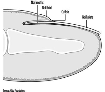
Figure 1 shows that the nail matrix is an invagination of the epithelium and it is covered by the nail fold at its base. A thin film of horny layer called the cuticle serves to seal the paronychial space by stretching from the nail fold to the nail plate.

Figure 1. The structure of the nail.

SKI040F1

The most vulnerable parts of the nail are the nail fold and the area beneath the tip of the nail plate, although the nail plate itself may suffer direct physical or chemical traumata. Chemical substances or infective agents may penetrate under the nail plate at its free margin. Moisture and alkali may destroy the cuticle and allow the entry of bacteria and fungi which will cause inflammation of the paronychial tissue and produce secondary growth disturbance of the nail plate.

The most frequent causes of nail disease are chronic paronychia, ringworm, trauma, psoriasis, impaired circulation and eczema or other dermatitis. Paronychia is an inflammation of the nail fold. Acute paronychia is a painful suppurative condition requiring antibiotic and sometimes surgical treatment. Chronic paronychia follows loss of the cuticle which allows water, bacteria and Candida albicans to penetrate into the paronychial space. It is common among persons with intense exposure to water, alkaline substances and detergents, such as kitchen staff, cleaners, fruit and vegetable preparers and canners and housewives. Full recovery cannot be achieved until the integrity of the cuticle and eponychium sealing the paronychial space has been restored.

Exposure to cement, lime and organic solvents, and work such as that of a butcher or poulterer may also cause trauma of the cuticle and nail folds.

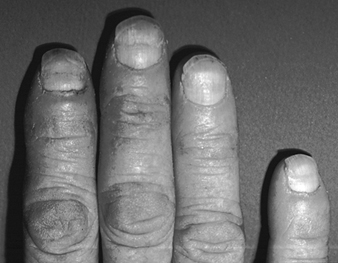
Any inflammation or disease of the nail matrix may result in dystrophy (distortion) of the nail plate, which is usually the symptom which has brought the condition to medical attention. Exposure to chilling cold, or the arterial spasm of Raynaud’s phenomenon, can also damage the matrix and produce nail dystrophy. Sometimes the damage is temporary and the nail dystrophy will disappear after removal of the cause and treatment of the inflammatory condition. (An example is shown in figure 2.)

Figure 2. Onychodystrophy secondary to contact dermatitis resulting from chronic irritation.

SKI040F2

One cause of nail damage is the direct application of certain cosmetic preparations, such as base coats under nail polish, nail hardeners and synthetic nail dressings to the nail.

Some special occupations may cause nail damage. There has been a report of dystrophy due to handling the concentrated dipyridylium pesticide compounds paraquat and diquat. During the manufacture of selenium dioxide, a fine powder of this substance may get under the fringe of the nail plate and cause intense irritation and necrosis of the finger tip and damage to the nail plate. Care should be taken to warn workers of this hazard and advise them always to clean the subungual areas of their fingers each day.

Certain types of allergic contact dermatitis of the finger tips frequently result in secondary nail dystrophy. Six common sensitizers which will do this are:

  1. amethocaine and chemically related local anaesthetics used by dental surgeons
  2. formalin used by mortuary attendants, anatomy, museum and laboratory assistants
  3. garlic and onion used by cooks
  4. tulip bulbs and flowers handled by horticulturists and florists
  5. p-tert-butylphenol formaldehyde resin used by shoe manufacturers and repairers
  6. aminoethylethanolamine used in some aluminium fluxes.


The diagnosis can be confirmed by a positive patch test. The condition of the skin and nails will recover when contact ceases.

Protective measures

In many cases nails can be safeguarded by the use of suitable hand protection. However, where hand exposure exists, nails should receive adequate care, consisting essentially of preserving the cuticle and protecting the subungual area. The skin under the free margin of the nails should be cleaned daily in order to remove foreign debris or chemical irritants. Where barrier creams or lotions are employed, care should be taken to ensure that the cuticle and the area under the free margin are coated.

To preserve the intact cuticle it is necessary to avoid excessive manicure or trauma, maceration by prolonged exposure to water, and dissolution by repeated exposure to alkali, solvent and detergent solutions.


Back

Read 9295 times Last modified on Tuesday, 11 October 2011 21:19

" DISCLAIMER: The ILO does not take responsibility for content presented on this web portal that is presented in any language other than English, which is the language used for the initial production and peer-review of original content. Certain statistics have not been updated since the production of the 4th edition of the Encyclopaedia (1998)."

Contents

Skin Diseases References

Adams, RM. 1988. Medicolegal aspects of occupational skin diseases. Dermatol Clin 6:121.

—. 1990. Occupational Skin Disease. 2nd edn. Philadelphia: Saunders.

Agner, T. 1991. Susceptibility of atopic dermatitis patients to irritant dermatitis caused by sodium lauryl sulfate. A Derm-Ven 71:296-300.

Balch, CM, AN Houghton, and L Peters. 1993. Cutaneous melanoma. In Cancer: Principles and Practice of Oncology, edited by VTJ DeVita, S Hellman, and SA Rosenberg. Philadelphia: JB Lippincott.

Beral, V, H Evans, H Shaw, and G Milton. 1982. Malignant melanoma and exposure to fluorescent lighting at work. Lancet II:290-293.

Berardinelli, SP. 1988. Prevention of occupational skin disease through use of chemical protective gloves. Dermatol Clin 6:115-119.

Bijan, S. 1993. Cancers of the skin. In Cancer: Principles & Practice of Oncology, edited by VTJ DeVita, S Hellman, and SA Rosenberg. Philadelphia: JB Lippincott.

Blair, A, S Hoar Zahm, NE Pearce, EF Heinerman, and J Fraumeni. 1992. Clues to cancer etiology from studies of farmers. Scand J Work Environ Health 18:209-215.

Commission de la santé et de la sécurité du travail. 1993. Statistiques sur les lesions professionnelles de 1989. Québec: CSST.

Cronin, E. 1987. Dermatitis of the hands in caterers. Contact Dermatitis 17: 265-269.

De Groot, AC. 1994. Patch Testing: Test Concentrations and Vehicles for 3,700 Allergens. 2nd ed. Amsterdam: Elsevier.

Durocher, LP. 1984. La protection de la peau en milieu de travail. Le Médecin du Québec 19:103-105.

—. 1995. Les gants de latex sont-ils sans risque? Le Médecin du Travail 30:25-27.

Durocher, LP and N Paquette. 1985. Les verrues multiples chez les travailleurs de l’alimentation. L’Union Médicale du Canada 115:642-646.

Ellwood, JM and HK Koh. 1994. Etiology, epidemiology, risk factors, and public health issues of melanoma. Curr Opin Oncol 6:179-187.

Gellin, GA. 1972. Occupational Dermatoses. Chicago: American Medical Assoc.

Guin, JD. 1995. Practical Contact Dermatitis. New York: McGraw-Hill.

Hagmar, L, K Linden, A Nilsson, B Norrving, B Akesson, A Schutz, and T Moller. 1992. Cancer incidence and mortality among Swedish Baltic Sea fisherman. Scand J Work Environ Health 18:217-224.

Hannaford, PC, L Villard Mackintosh, MP Vessey, and CR Kay. 1991. Oral contraceptives and malignant melanoma. Br J Cancer 63:430-433.

Higginson, J, CS Muir, and M Munoz. 1992. Human Cancer: Epidemiology and Environmental
Causes. Cambridge Monographs on Cancer Research. Cambridge, UK: CUP.

International Agency for Research on Cancer (IARC). 1983. Polynuclear aromatic compounds, Part I, Chemical, environmental and experimental data. Monographs on the Evaluation of the Carcinogenic Risk of Chemicals to Humans, No. 32. Lyon: IARC.

—. 1984a. Polynuclear aromatic compounds, Part 2, Carbon blacks, mineral oils and some Nitroarenes. Monographs on the Evaluation of the Carcinogenic Risk of Chemicals to Humans, No. 33. Lyon: IARC.

—. 1984b. Polynuclear aromatic compounds, Part 3, Industrial exposures in aluminium production, coal gasification, coke production, and iron and steel founding. Monographs on the Evaluation of the Carcinogenic Risk of Chemicals to Humans, No. 34. Lyon: IARC.

—. 1985a. Polynuclear aromatic compounds, Part 4, Bitumens, coal tars and derived products, shale-oils and soots. Monographs on the Evaluation of the Carcinogenic Risk of Chemicals to Humans, No. 35. Lyon: IARC.

—. 1985b. Solar and ultraviolet radiation. Monographs on the Evaluation of the Carcinogenic Risk of Chemicals to Humans, No. 55. Lyon: IARC.

—. 1987. Overall Evaluations of Carcinogenecity: An updating of IARC Monographs Volumes 1 to 42. Monographs on the Carcinogenic Risks to Humans. Suppl. 7. Lyon: IARC

—. 1990. Cancer: Causes, occurrence and control. IARC Scientific Publications, No. 100. Lyon: IARC.

—. 1992a. Cancer incidence in five continents. Vol. VI. IARC Scientific Publications, No. 120. Lyon: IARC.

—. 1992b. Solar and ultraviolet radiation. Monographs On the Evaluation of Carcinogenic Risks to Humans, No. 55. Lyon: IARC.

—. 1993. Trends in cancer incidence and mortality. IARC Scientific Publications, No. 121. Lyon: IARC.

Koh, HK, TH Sinks, AC Geller, DR Miller, and RA Lew. 1993. Etiology of melanoma. Cancer Treat Res 65:1-28.

Kricker, A, BK Armstrong, ME Jones, and RC Burton. 1993. Health, solar UV radiation and environmental change. IARC Technical Report, No. 13. Lyon: IARC.

Lachapelle, JM, P Frimat, D Tennstedt, and G Ducombs. 1992. Dermatologie professionnelle et de l’environnement. Paris: Masson.

Mathias, T. 1987. Prevention of occupational contact dermatitis. J Am Acad Dermatol 23:742-748.

Miller, D and MA Weinstock. 1994. Nonmelanoma skin cancer in the United States: Incidence. J Am Acad Dermatol 30:774-778.

Nelemans, PJ, R Scholte, H Groenendal, LA Kiemeney, FH Rampen, DJ Ruiter, and AL Verbeek. 1993. Melanoma and occupation: results of a case-control study in The Netherlands. Brit J Ind Med 50:642-646.

Rietschel, RI, and JF Fowler Jr. 1995. Fisher’s Contact Dermatitis. 4th ed. Baltimore: Williams & Wilkins.

Sahel, JA, JD Earl, and DM Albert. 1993. Intraocular melanomas. In Cancer: Principles & Practice of Oncology, edited by VTJ DeVita, S Hellman, and SA Rosenberg. Philadelphia: JB Lippincott.

Sasseville, D. 1995. Occupational dermatoses: Employing good diagnostic skills. Allergy 8:16-24.

Schubert, H, N Berova, A Czernielewski, E Hegyi and L Jirasek. 1987. Epidemiology of nickel allergy. Contact Dermatitis 16:122-128.

Siemiatycki J, M Gerin, R Dewar, L Nadon, R Lakhani, D Begin, and L Richardson. 1991. Associations between occupational circumstances and cancer. In Risk Factors for Cancer in the Workplace, edited by J Siematycki. London, Boca Raton: CRC Press.

Stidham, KR, JL Johnson, and HF Seigler. 1994. Survival superiority of females with melanoma. A multivariate analysis of 6383 patients exploring the significance of gender in prognostic outcome. Archives of Surgery 129:316-324.

Turjanmaa, K. 1987. Incidence of immediate allergy to latex gloves in hospital personnel. Contact Dermatitis 17:270-275.