Tuesday, 08 March 2011 15:49

Stigmata

Rate this item
(0 votes)

Occupational stigma or occupational marks are work-induced anatomical lesions which do not impair working capacity. Stigmata are generally caused by mechanical, chemical or thermal skin irritation over a long period and are often characteristic of a particular occupation. Any kind of pressure or friction on the skin may produce an irritating effect, and a single violent pressure may break the epidermis, leading to the formation of excoriations, seropurulent blisters and infection of the skin and underlying tissues. On the other hand, however, frequent repetition of moderate irritant action does not disrupt the skin but stimulates defensive reactions (thickening and keratinization of the epidermis). The process may take three forms:

  1. a diffuse thickening of the epidermis which merges into the normal skin, with preservation and occasional accentuation of skin ridges and unimpaired sensitivity
  2. a circumscribed callosity made up of smooth, elevated, yellowish, horny lamellae, with partial or complete loss of skin ridges and impairment of sensitivity. The lamellae are not circumscribed; they are thicker in the centre and thinner towards the periphery and blend into the normal skin
  3. a circumscribed callosity, mostly raised above the normal skin, 15 mm in diameter, yellowish-brown to black in colour, painless and occasionally associated with hypersecretion of the sweat glands.


Callosities are usually produced by mechanical agents, sometimes with the aid of a thermal irritant (as in the case of glass- blowers, bakers, firefighters, meat curers, etc.), when they are dark-brown to black in colour with painful fissures. If, however, the mechanical or thermal agent is combined with a chemical irritant, callosities undergo discoloration, softening and ulceration.

Callosities which represent a characteristic occupational reaction (particularly on the skin of the hand as shown in figure 1 and 2) are seen in many occupations. Their form and localization are determined by the site, force, manner and frequency of the pressure exerted, as well as by the tools or materials used. The size of callosities may also reveal a congenital tendency to skin keratinization (ichthyosis, hereditary keratosis palmaris). These factors may also often be decisive as concerns deviations in the localization and size of callosities in manual workers.

Figure 1. Occupational stigmata on the hands.

SKI050F1

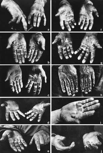
(a) Tanner’s ulcers; (b) Blacksmith; (c) Sawmill worker; (d) Stonemason; (e) Mason; (f) Marble Mason; (g) Chemical factory worker; (h) Paraffin refinery worker; (I) Printer; (j) Violinist 

 (Photos: Janina Mierzecka.)

Figure 2. Calluses at pressure points on the palm of the hand.

SKI050F2

Callosities normally act as protective mechanisms but may, under certain conditions, acquire pathological features; for this reason they should not be overlooked when pathogenesis and, particularly, prophylaxis of occupational dermatoses are envisaged.

When a worker gives up a callosity-inducing job, the superfluous horny layers undergo exfoliation, the skin becomes thin and soft, the discoloration disappears and the normal appearance is restored. The time required for skin regeneration varies: occupational callosities on the hands may occasionally be seen several months or years after the work has been given up (especially in blacksmiths, glass-blowers and sawmill workers). They persist longer in senile skin and when associated with connective tissue degeneration and bursitis.

Fissures and erosions of the skin are characteristic of certain occupations (railway workers, gunsmiths, bricklayers, goldsmiths, basket weavers, etc.). The painful “tanner’s ulcer” associated with chromium compound exposures (figure 1) round or oval in shape and from 2-10 mm in diameter. The localisation of occupational lesions (e.g. on confectioners’ fingers, tailors’ fingers and palms, etc.) is also characteristic.

Pigment spots are caused by the absorption of dyes through the skin, the penetration of particles of solid chemical compounds or industrial metals, or the excessive accumulation of the skin pigment, melanin, in workers in coking or generator plants, after three to five years of work. In some establishments, about 32% of workers were found to exhibit melanomata. Pigment spots are mostly found in chemical workers.

As a rule, dyes absorbed through the skin cannot be removed by routine washing, hence their permanence and significance as occupational stigmata. Pigment spots occasionally result from impregnation with chemical compounds, plants, soil or other substances to which the skin is exposed during the work process.

A number of occupational stigmata may be seen in the region of the mouth (e.g. Burton’s line within the gums of workers exposed to lead, teeth erosion in workers exposed to acid fumes, etc. blue colouring of the lips in workers engaged in aniline manufacture and in the form of acne. Characteristic odours connected with certain occupations may also be considered as occupational stigmata.


Back

Read 9906 times Last modified on Tuesday, 11 October 2011 21:06
More in this category: « Occupational Nail Dystrophy

" DISCLAIMER: The ILO does not take responsibility for content presented on this web portal that is presented in any language other than English, which is the language used for the initial production and peer-review of original content. Certain statistics have not been updated since the production of the 4th edition of the Encyclopaedia (1998)."

Contents

Skin Diseases References

Adams, RM. 1988. Medicolegal aspects of occupational skin diseases. Dermatol Clin 6:121.

—. 1990. Occupational Skin Disease. 2nd edn. Philadelphia: Saunders.

Agner, T. 1991. Susceptibility of atopic dermatitis patients to irritant dermatitis caused by sodium lauryl sulfate. A Derm-Ven 71:296-300.

Balch, CM, AN Houghton, and L Peters. 1993. Cutaneous melanoma. In Cancer: Principles and Practice of Oncology, edited by VTJ DeVita, S Hellman, and SA Rosenberg. Philadelphia: JB Lippincott.

Beral, V, H Evans, H Shaw, and G Milton. 1982. Malignant melanoma and exposure to fluorescent lighting at work. Lancet II:290-293.

Berardinelli, SP. 1988. Prevention of occupational skin disease through use of chemical protective gloves. Dermatol Clin 6:115-119.

Bijan, S. 1993. Cancers of the skin. In Cancer: Principles & Practice of Oncology, edited by VTJ DeVita, S Hellman, and SA Rosenberg. Philadelphia: JB Lippincott.

Blair, A, S Hoar Zahm, NE Pearce, EF Heinerman, and J Fraumeni. 1992. Clues to cancer etiology from studies of farmers. Scand J Work Environ Health 18:209-215.

Commission de la santé et de la sécurité du travail. 1993. Statistiques sur les lesions professionnelles de 1989. Québec: CSST.

Cronin, E. 1987. Dermatitis of the hands in caterers. Contact Dermatitis 17: 265-269.

De Groot, AC. 1994. Patch Testing: Test Concentrations and Vehicles for 3,700 Allergens. 2nd ed. Amsterdam: Elsevier.

Durocher, LP. 1984. La protection de la peau en milieu de travail. Le Médecin du Québec 19:103-105.

—. 1995. Les gants de latex sont-ils sans risque? Le Médecin du Travail 30:25-27.

Durocher, LP and N Paquette. 1985. Les verrues multiples chez les travailleurs de l’alimentation. L’Union Médicale du Canada 115:642-646.

Ellwood, JM and HK Koh. 1994. Etiology, epidemiology, risk factors, and public health issues of melanoma. Curr Opin Oncol 6:179-187.

Gellin, GA. 1972. Occupational Dermatoses. Chicago: American Medical Assoc.

Guin, JD. 1995. Practical Contact Dermatitis. New York: McGraw-Hill.

Hagmar, L, K Linden, A Nilsson, B Norrving, B Akesson, A Schutz, and T Moller. 1992. Cancer incidence and mortality among Swedish Baltic Sea fisherman. Scand J Work Environ Health 18:217-224.

Hannaford, PC, L Villard Mackintosh, MP Vessey, and CR Kay. 1991. Oral contraceptives and malignant melanoma. Br J Cancer 63:430-433.

Higginson, J, CS Muir, and M Munoz. 1992. Human Cancer: Epidemiology and Environmental
Causes. Cambridge Monographs on Cancer Research. Cambridge, UK: CUP.

International Agency for Research on Cancer (IARC). 1983. Polynuclear aromatic compounds, Part I, Chemical, environmental and experimental data. Monographs on the Evaluation of the Carcinogenic Risk of Chemicals to Humans, No. 32. Lyon: IARC.

—. 1984a. Polynuclear aromatic compounds, Part 2, Carbon blacks, mineral oils and some Nitroarenes. Monographs on the Evaluation of the Carcinogenic Risk of Chemicals to Humans, No. 33. Lyon: IARC.

—. 1984b. Polynuclear aromatic compounds, Part 3, Industrial exposures in aluminium production, coal gasification, coke production, and iron and steel founding. Monographs on the Evaluation of the Carcinogenic Risk of Chemicals to Humans, No. 34. Lyon: IARC.

—. 1985a. Polynuclear aromatic compounds, Part 4, Bitumens, coal tars and derived products, shale-oils and soots. Monographs on the Evaluation of the Carcinogenic Risk of Chemicals to Humans, No. 35. Lyon: IARC.

—. 1985b. Solar and ultraviolet radiation. Monographs on the Evaluation of the Carcinogenic Risk of Chemicals to Humans, No. 55. Lyon: IARC.

—. 1987. Overall Evaluations of Carcinogenecity: An updating of IARC Monographs Volumes 1 to 42. Monographs on the Carcinogenic Risks to Humans. Suppl. 7. Lyon: IARC

—. 1990. Cancer: Causes, occurrence and control. IARC Scientific Publications, No. 100. Lyon: IARC.

—. 1992a. Cancer incidence in five continents. Vol. VI. IARC Scientific Publications, No. 120. Lyon: IARC.

—. 1992b. Solar and ultraviolet radiation. Monographs On the Evaluation of Carcinogenic Risks to Humans, No. 55. Lyon: IARC.

—. 1993. Trends in cancer incidence and mortality. IARC Scientific Publications, No. 121. Lyon: IARC.

Koh, HK, TH Sinks, AC Geller, DR Miller, and RA Lew. 1993. Etiology of melanoma. Cancer Treat Res 65:1-28.

Kricker, A, BK Armstrong, ME Jones, and RC Burton. 1993. Health, solar UV radiation and environmental change. IARC Technical Report, No. 13. Lyon: IARC.

Lachapelle, JM, P Frimat, D Tennstedt, and G Ducombs. 1992. Dermatologie professionnelle et de l’environnement. Paris: Masson.

Mathias, T. 1987. Prevention of occupational contact dermatitis. J Am Acad Dermatol 23:742-748.

Miller, D and MA Weinstock. 1994. Nonmelanoma skin cancer in the United States: Incidence. J Am Acad Dermatol 30:774-778.

Nelemans, PJ, R Scholte, H Groenendal, LA Kiemeney, FH Rampen, DJ Ruiter, and AL Verbeek. 1993. Melanoma and occupation: results of a case-control study in The Netherlands. Brit J Ind Med 50:642-646.

Rietschel, RI, and JF Fowler Jr. 1995. Fisher’s Contact Dermatitis. 4th ed. Baltimore: Williams & Wilkins.

Sahel, JA, JD Earl, and DM Albert. 1993. Intraocular melanomas. In Cancer: Principles & Practice of Oncology, edited by VTJ DeVita, S Hellman, and SA Rosenberg. Philadelphia: JB Lippincott.

Sasseville, D. 1995. Occupational dermatoses: Employing good diagnostic skills. Allergy 8:16-24.

Schubert, H, N Berova, A Czernielewski, E Hegyi and L Jirasek. 1987. Epidemiology of nickel allergy. Contact Dermatitis 16:122-128.

Siemiatycki J, M Gerin, R Dewar, L Nadon, R Lakhani, D Begin, and L Richardson. 1991. Associations between occupational circumstances and cancer. In Risk Factors for Cancer in the Workplace, edited by J Siematycki. London, Boca Raton: CRC Press.

Stidham, KR, JL Johnson, and HF Seigler. 1994. Survival superiority of females with melanoma. A multivariate analysis of 6383 patients exploring the significance of gender in prognostic outcome. Archives of Surgery 129:316-324.

Turjanmaa, K. 1987. Incidence of immediate allergy to latex gloves in hospital personnel. Contact Dermatitis 17:270-275.